- Volume 60 , Number 2
- Page: 277–8
Improved method for purification of Mycobacterium leprae f rom armadillo tissues
To the Editor:
An efficient, rapid, and cheap method for the preparation of pure Mycobacterium leprae from armadillo tissues is described. It is especially recommended for application in immunology and molecular genetics.
Because the cultivation of M . leprae has not yet been possible, the only source for specified living M . leprae are animal models, especially the nine-banded armadillo (Dasypus novemcintus). Difficulty in the separation and purification of the organism from armadillo tissues is one of the main problems workers face in this field, and it limits the effective use of M . leprae in immunology, vaccine preparation, biochemical studies, etc.
We have developed a rapid, efficient, cheap, and easy method for the preparation of M. leprae from armadillo tissues which provides bacteria of high quantity and high quality. The method depends on low speed/ high speed centrifugation with the use of Percoll as a gradient separation medium.
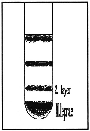
Armadillo liver tissue (6 grams) from an armadillo inoculated with M. leprae 24 months earlier was homogenized with ultraturax at high speed in 10 ml phosphate buffered saline (PBS), pH 7.0, in a Beckman screw-cap centrifuge tube (Cat. no. 355670), and the homogenate was then centrifuged at 55 x g for 6 min at 5ºC. The supernatant was carefully aliquoted to two or three 5-ml screw-cap tubes (Greiner, Cat. no. 124261) and centrifuged at 15,000 x g for 10 min at 5ºC. The supernatant was discarded, and the sediment was resuspended in 6 ml Tween buffer [1 ml 10% Tween 80/100 ml distilled water/0.2 ml of 2-morpholino-ethane-sulfonic acid (MES) (1.0661 g/10 ml distilled water, pH 6.8, adjusted by NaOH)].' The suspensions were pooled in a sterile Beckman screw-cap centrifuge tube (as above), and 24 ml of 30% Percoll [30 ml Percoll/ 70 ml Tween buffer (as described above)] was added. The tube was centrifuged at 70,000 x g for 1 hr at 5ºC. The bottom layer (The Figure) was taken and washed twice with Tween buffer, twice with saline solution, and twice with PBS at 13,000 x g for 10 min at 5ºC. The sediment was resuspended in 2 ml PBS and kept for further use.

The figure. Separations of M. leprae suspensionsafter centrifugation.
The present method takes about 2 ½ hr and provides M . leprae free of any armadillo tissue contaminations. The number of viable organisms is very high. The second layer (The Figure) can be used for methods with lesser needs for purity of the bacteria. The count in this fraction lies two powers below the bottom layer, and it is contaminated with armadillo tissue cells.
The standard protocol for purification of M. leprae described by Draper (1) is timeconsuming compared to this new method. The efficiency of our method and the viability of the bacteria prepared with it are at least as high as those with the Draper protocol. The bacteria prepared by the present protocol were white, i.e., devoid of a ferric iron-protein complex which makes the use of the bacteria unacceptable in immunological studies in man (2, 3).
- Cornelia Rodde
Abdel-Hamid A. F. Mohamed, M.Sc.
Hans Gerd Liiesse, Dipl.-Ing. agr.
Jindrich Kazda, M.V.D., Ph.D.
Research Institute for Experimental Biology and Medicine
Parkallee 1-40
D-2061 Borstel, Germany
REFERENCES
1. DRAPER, P. Protocol 1/79, Purification of M. leprae. In: Report on the Enlarged SC Meeting, Geneva, 7-8 February 1979. Annex 1. Geneva: World Health Organization, 1980. TDR/IMMLEP-SWG
2. SHEPARD, C. C., DRAPER, P., REES, R. J. W. and LOWE, C. Effect of purification steps on the immunogenicity of M. leprae. Br. J. Pathol. 61(1980)376-379.
3. SPECIAL PROGRAMME for RESEARCH and TRAINING in TROPICAL DISEASES. Sixth programme report. Geneva: World Health Organization, 1983, pp. 220-80.3. 221. TDR/PR-6/83.8-LEP.
Reprint requests to Professor Kazda.