- Volume 60 , Number 3
- Page: 483–5
Unusual cell of leprous neuritis
To The Editor:
For the past two decades we have been studying leprous nerve lesions from various angles, and the use of electron microscopy has proved particularly useful in defining some of the early as well as aberrent pathology (4,5). In this spectral disease the exact identification of a battery of infiltrating lymphoid and phagocytic cells, seen in different stages of development, is very difficult at the fine structural level. However, every cell is an entity by itself with a morphological feature of its own, and it is here that electron microscopy helps in elucidating the details.
In this short communication we wish to report on the presence of an unusual cell type frequently seen in active and chronic nerve lesions obtained from both treated and untreated cases of borderline (BB) to lepromatous (LL) leprosy (3) and as yet unreported.
The cells in question were seen within the endoneurium, were strikingly rich in rough endoplasmic reticulum (RER), and were seen in close association with small groups of axons and Schwann cells. Moreover, these cells were seen with an even or sometimes irregular coating of basal lamina around them. The stacking of RER and the nuclear appearance of some of the cells resembled that of a plasma cell (Fig. 1). We have not seen any presence of M. leprae in these unusual cells, but a bacterial presence was seen in the Schwann cell processes lying in close association with them.
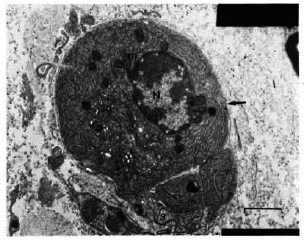
Fig. 1. Electron micrograph of one of the unusual cells from part of a sural nerve biopsy obtained from a multidrug-treated BL patient. There are abundant arrays of rough endoplasmic reticulum and a well-developed golgi complex (g) within the cytoplasm. Mitochondria (m) and a few dense bodies are also seen. A small part of the cell cytoplasm resembles that of a Schwann cell(s). Note the even coating of the basal lamina (arrow) around this cell. N = nucleus. Uranyl acetate and lead citrate stain; bar = 1 µm.
The frequency with which these cells were seen in several of the nerve lesions, to our mind, does not appear to be a chance finding. However, the origin and function of these cells is debatable. They could be either transformed Schwann cells or migratory cells that have invaded the basal laminal tube of the Schwann cells. That Schwann cells are known to synthesize and to secrete collagen
and increase in endoplasmic reticulum (both smooth and rough) has been reported in activated Schwann cells (1) but not to the extent and type that has been seen in these unusual cells.
One strong evidence that these are probably migratory cells that have invaded the basal laminal tubes of the Schwann cells is the presence of an occasional foot-print-like process seen protruding from the basal laminal tube (Fig. 2).
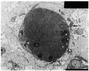
Fig. 2. Electron micrograph of another unusual cell from a sural nerve biopsy from a multidrug-treated BL patient showing foot-print-like processes (arrow) protruding out of the basal laminal tube. The continuity of the basal lamina is disrupted at these sites. Note the presence of abundant arrays of rough endoplasmic reticulum and an axon-like profile with an extended axolemma (arrowhead) on one side. Uranyl acetate and lead citrate stain; bar = 1 µm
One of the cell types known to invade the basal laminal tube of Schwann cells are activated macrophages. In a demyelinating disorder like Guillain-Barré syndrome (GBS) and its experimental counterpart experimental allergic neuritis (EAN), macrophages invade the basal laminal tube and take an active role in stripping, phagocytosis, and breakdown of myelin in an immune-mediated process (2). However, the morphological appearance of the cells in question does not resemble an activated macrophage nor were they ever seen in the process of engulfing myelin.
Other known cells which are rich in RER are the collagen-synthesizing fibroblasts, immunoglobulin-secreting plasma cells, and the secretory epithelioid cells that have been described in granulomas of delayed-type hypersensitivity reactions (6). None of these cells are known phagocytes and, if it can be assumed that any one of these cell types could have invaded (or become trapped in) the basal laminal tube of a Schwann cell, then what function they subserve remains an open question.
- V . P. Shetty, Ph.D.
Research Officer
- N . H. Antia, F.R.C.S., F.A.C.S. (Hon.)
Director and Trustee
The Foundation for Medical Research
84-A R. G. Thadani Marg. Worli
Bombay 400018, India
Acknowledgment. We thank Miss Suchitra K. for technical assistance, Mr. Harish Poojary for the electron micrographs, and Mr. A. Venugopalan for careful typing.
REFERENCES
1. ASBURY, K. A. The biology of Schwann cells. In: Peripheral Neuropathy. Vol. 1. Dyck, P.J., Thomas, P.K. and Lambert, W., eds., Philadelphia: W.B. Sanders Company, 1975, pg. 201-212.
2. RAINE, C. S. Experimental allergic encephalomyelitis and experimental allergic neuritis. In: Handbook of Clinical Neurology. P.J. Vinken, et al., eds. Amsterdam: Elsevier. 47(1985)429-466.
3. RIDLEY, D. S. and JOPLING, W. H. Classification and of leprosy according to immunity; a five-group system. Int. J. Lepr. 34(1966)255-273.
4. SHETTY, V. P. and ANTIA, N. H. Degeneration and regeneration of unmyelinated fibres in experimental leprous neuropathy. Int. J. Lepr. 49(1981)324-330.
5. SHETTY, V. P., ANTIA, N. H. and JACOBS, J. M. The pathology of early. J. Neurol. Sci. 88(1988)115-131.
6. TURK, L. J. and NARAYAN, R. B. The origin, morphology and function of epithelioid cells. Immuleprous neuropathy.- nobiology 161(1982)274-282.
Reprint requests to Dr. Antia