- Volume 58 , Number 2
- Page: 392–3
Role of S-100 protein as a marker for schwann cells in the diagnosis of tuberculoid leprosy
To the Editor:
The histopathological diagnosis of leprosy, especially in its early stages, is by no means easy. An unequivocal diagnosis can be made only when acid-fast organisms are found in a dermal nerve in the lesion. This is often not possible, especially in the lesions of tuberculoid leprosy in which Mycobacterium leprae are extremely rare. Active destruction of a nerve by granulomatous inflammation is an equally reliable finding to confirm the diagnosis of tuberculoid leprosy. However, once in a while we come across skin biopsies with granulomatous inflammation with no identifiable nerves. Several focal granulomas, both in the dermis and the subcutaneous tissue which might have been " graveyards" of nerves, are seen; but the remnants of nerve left behind are too fragmented to be identified with certainty in the hematoxylin and eosin-stained sections or in sections stained for acid-fast organisms.
Granulomatous lesions of the skin, such as sarcoidosis, tuberculosis, tuberculids, tertiary syphilis, deep mycotic infections, etc., are so similar to tuberculoid leprosy that evidence of nerve destruction is a significant finding for differentiating it from other granulomatous lesions.
In an exhaustive study of the distribution of the S-100 protein in normal tissues using immunoperoxidase reaction, its presence was reported in melanocytes, Langerhans' cells, ducts of salivary and sweat glands, serous glands of the lung, sustentacular cells of the adrenal, reticulum cells of the lymph node, chondrocytes, myoepithelial cells, glial cells and Schwann cells (2). Fleury and Bacchi in an elegant study were able to visualize peripheral nerves in tuberculoid granulomas by staining for the S-100 protein using the immunoperoxidase reaction (1). In this communication, we record our experiences in using the S-100 protein as a marker for Schwann cells in the diagnosis of tuberculoid leprosy.
In our histopathological examinations for the diagnosis and classification of leprosy, several sections stained with hematoxylin and eosin and a modified Fite's stain for acid-fast bacilli are routinely examined. During a 5-year period beginning in 1984, there were 20 skin biopsies in our files reported as granulomatous inflammation of the skin in which a definite diagnosis of tuberculoid leprosy could not be made because of our inability to demonstrate destruction of dermal nerves by the granuloma. As controls, biopsies of five patients diagnosed as sarcoidosis of the skin and of two patients with skin tuberculosis were also chosen from our files. Four μm-thick sections were prepared from the paraffin blocks of these 27 biopsies. All of the sections were processed as follows for staining the S-100 protein.
The sections were deparallinized in xylene, hydrated in graded ethanol concentrations to distilled water, overlaid with 1% hydrogen peroxide for 15 min to block endogenous peroxidases, and washed with distilled water and phosphate buffered saline (PBS) (pH 7.4). The sections were then treated with normal goat serum at a 1.5:100 dilution for 20 min. and the excess blotted away. All further incubations were done at 37ºC in a humidified chamber. The sections were treated with primary antiserum consisting of rabbit antiserum to bovine S-100 protein (Dakopatts, Santa Barbara, California, U.S.A.) at a 1:2000 dilution for 2.5 hr. For a negative control, the sections were overlaid with only PBS. They were then treated for 30 min with biotinylaled goat antiserum to rabbit immunoglobulin (Vectastain; Vector Laboratories, Burlingame, California, U.S.A.) at a 1:200 dilution, washed with PBS, and incubated with peroxidase-conjugated avidin-biotin complex in a 1:50 dilution (Vectastain) for 30 min. Finally, they were overlaid for 5 min with 0.025% 3'diaminobenzidine (Polysciences, Inc., Warrington, Pennsylvania, U.S.A.) in a PBS buffer, pH 7.4, and activated with hydrogen peroxide to bring up the brown color. The sections were then counter-stained with hematoxylin, dehydrated in graded ethanol concentrations, cleared in xylene, and mounted in Richard Allan mounting medium.
In 14 of the 20 biopsies under investigation, dark staining fragments of tissue morphologically identifiable as portions of peripheral nerves were found (The Figure). There were both longitudinal and transverse sections of nerve fragments clearly identifiable in the granuloma. These were obviously remnants of nerves which had undergone destruction by the granulomatous inflammation and, therefore, a diagnosis of tuberculoid leprosy could be made on these 14 patients. Well-preserved and intact longitudinal and cross-sections of nerves, some in the middle of the granulomas, were seen in all five biopsies of sarcoidosis and in two biopsies of tuberculosis. These small intact nerves could be differentiated from nerves that had undergone fragmentation in the granulomas of tuberculoid leprosy. In addition, melanocytes and Langerhans' cells in the epidermis, dendritic cells in the granulomas, and sweat ducts in the dermis were also stained. The dendritic cells and the sweat ducts in the granuloma were easily differentiated from nerve fragments by their morphology.
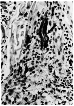
The Figure. Section clearly showing fragments of peripheral nerves (immunoperoxidase stain for S-100 x 250).
Of these 14 biopsies from which a diagnosis of tuberculoid leprosy was confirmed, 7 belonged to the polar tuberculoid group and 7 to the borderline tuberculoid group. We strongly recommend the use of the S-100 protein as a marker to identify peripheral nerves to aid in the histopathological diagnosis of tuberculoid leprosy lesions.
- Charles K. Job, M.D., F.R.C.Path.
Vernia Drain, M.D.
Angelina T. Doming, B.S.
Robert C. Hastings, M.D., Ph.D.
Laboratory Research Branch
GWL Hansen's Disease Center
Carville, Louisiana 70721, U.S.A.
- Michael A. Gerber, M.D., Ph.D.
Department of Pathology
Tulane University Medical School
New Orleans, Louisiana, U.S.A.
REFERENCES
1. Fleury. R. N. and Bacchi, E. E. S-100 protein and immunoperoxidase technique as an aid in the histopathologic diagnosis of leprosy. Int. J. Lepr. 55(1987)338-344.
2. Kahn, H., Marks, A., Thom. II. and Baumel, R. Role of antibody S-100 protein in diagnostic pathology. Am. J. Clin. Pathol. 79(1983)341-347.