- Volume 57 , Number 4
- Page: 873–4
Aggregation phenomenon of cultivated blood macrophages isolated f rom leprosy patients
To the Editor:
We report on the aggregation phenomenon of cultivated blood macrophages derived from leprosy patients. Macrophages that were isolated formed aggregates over a coverslip (Fig. 1) in almost all of the patients with progressive stages of leprosy, such as new patients, many patients with erythema nodosum leprosum (ENL), and those having a relapse and leprous neuralgia. On the other hand, macrophages isolated from patients with leprosy in the quiescent stages were almost always dispersed (Fig. 2). Thirteen of the 16 patients whose cells formed aggregates were in a progressive stage of the disease and 17 of the 20 patients whose cells were dispersed had quiescent leprosy. As described later, the blood macrophages were isolated from leukocytes in supernatant plasma following red cell precipitation. However, as is well known, no plasma can be isolated from healthy individuals through this method. Thus, we could not use healthy subjects as controls. The differences between the two patient groups were statistically significant (p < 0.01, x2).
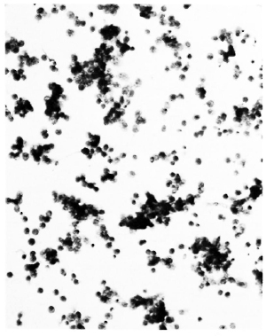
Fig. 1. Cell aggregate form of macrophages isolated from a patient in a progressive stage of leprosy ( x 100).
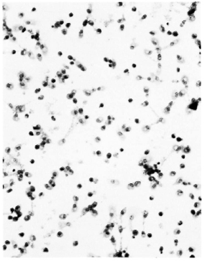
Fig. 2. Cell dispersion form of macrophages isolated from a patient in a quiescent stage of leprosy (x 100).
Subjects used in this study were randomly selected from among patients treated at the National Leprosarium Tama-Zensyo-En, without regard to age, sex, type or stage of leprosy. Fifteen ml of heparinized blood was collected from each patient using a 30-ml plastic syringe. After collection, the blood stood for 1 hr at room temperature; the red cells were precipitated; and approximately 7 ml plasma was harvested as a supernatant in which almost all of the monocytes were floating. The plasma was mixed with penicillin 100 U/ml, and approximately 1 ml of the plasma was poured into a plastic Leighton tube (Costar Co., Cambridge, Massachusetts, U.S.A.) with a Lux Ther-manox coverslip (Miles Laboratories, Inc., Naperville, Illinois, U.S.A.). and cultivated in a CO2 incubator at 37ºC. Each coverslip was removed 3 days after cultivation, fixed with methanol, and stained with Giemsa's solution.
There are a number of reports thought to be related to our study (1-6). However, we see no direct relation between these findings and ours, nor are there reports suggesting that macrophages in leprous plasma react with something during cultivation. The nature of this cell aggregation is such that we think the macrophages could be reacting with antigens in the plasma of leprosy patients, and forming a granuloma in vitro. These results indicate that cell aggregation can be used as an indicator of the progressive stages of leprosy.
- A. Yamagami, M.D.
Chief, Bacteriological Laboratory
National Institute for Leprosy Research
Aoba-cho, Higashi-Murayama
Tokyo 189, Japan
- M. Koseki, M.D.
Chief, Dermatology Section
- S. Kim, M.D.
S. Hazama, M.D.
K. Sanada, M.D.
Dermatologists
National Leprosarium Tama-Zensyo-En
Aoba-cho, Higashi-Murayama
Tokyo 189, Japan
REFERENCES
1. Azulay, R. D. Chemotaxis of monocytes in hanseniasis. Int. J. Lepr. 50(1982)215-216.
2. Fuquay, J. I., Loo, D. T. and Barnes, D. W. Binding of Staphylococcus aureus by human serum spreading factor in an in vitro assay. Infect. Immun. 52(1986)714-717.
3. Jones, G. E., Pizzey, J. A. and Witkowski, A. The effect of monensin on cell aggregation of normal and dystrophic human skin fibroblasts. Exp. Cell Res. 159(1985)540-545.
4. Laal, S., Bhutani. L. K. and Nath, I. Natural emergence of antigen-reactive T cells in lepromatous leprosy patients during erythema nodosum leprosum. Infect. Immun. 50(1985)887-892.
5. Masuda, A. and Scheinderg, M. A. Peripheral blood monocyte function in leprosy patients. Int. J. Lepr. 48(1980)254-259.
6. Weisbart, R. H., Golde, D. W., Spolter, L., Eggena, P. and Rinderknecht, H. Neutrophil migration inhibition factor from T lymphocytes (NIF-T): a new lymphokine. Clin. Immunol. Immunopathol. 14(1979)441-448.