- Volume 58 , Number 1
- Page: 73–77
Analysis of variation in batches of armadillo-derived Mycobacterium leprae by immunoblotting
ABSTRACT
Several batches of cell-free extracts of armadillo-derived Mycobacterium leprae were analyzed by SDS-PAGE and by immunoblotting with monoclonal antibodies. The presence or absence of protease inhibitors had a profound effect on the protein antigens, particularly the 65-kDa antigen. In the absence of protease inhibitors, there were both quantitative and qualitative differences between the different batches of M. leprae extracts.
RÉSUMÉ
Plusieurs lots d'extraits sans cellule de Mycobacterium leprae obtenus chez le tatou ont été analyses par la méthode SDS-PAGE, et par l'"immunoblotting" avec des anticorps monoclonaux. La présence ou l'absence d'inhibiteurs de la protéase avaient un effet prononcé sur les antigènes protéiniques, et particulièrement sur l'antigène 65-kDa. En l'absence d'inhibiteurs de la protease, on a observé des differences à la fois quantitatives et qualitatives entre les différents lots des extraits de M. leprae.
RESUMEN
Se prepararon varios lotes de extractos de Mycobacterium leprae aislados de armadillos, se fraccionaron por clectroforesis en gel de poliacrilamida (SDS-PAGE) e inmunoelectrotransferencia, y se hicieron reaccionar con anticuerpos monoclonales. La presencia o ausencia de inhibidores de proteasas tuvo un profundo efecto sobre los antígenos proteicos, principalmente sobre el antígeno de 65 kDa. En ausencia de inhibidores de proteasas hubieron diferencias cuali-y cuantitativas entre los diferentes lotes de los extractos de M. leprae.
Studies of the immunochemistry of Mycobacterium leprae, the etiologic agent of leprosy, have been impeded by the inability to cultivate the bacillus on artificial medium. The discovery that nine-banded armadillos were susceptible to infection with M. leprae (6) was of paramount importance since armadillo-infected tissues made an excellent source of large amounts of bacteria. This material has now been used in large numbers of laboratories in a variety of immunological and biochemical studies. A number of groups have studied the antibody responses to M. leprae using anti-M. leprae antisera, leprosy patients' sera, and monoclonal antibodies (1, 2, 4, 7, 10). However, both the number of M. leprae components revealed and their molecular weight range varied considerably between different studies. It is possible that these differences were due to variations in techniques and the sera used. However, the possibility that major differences can occur between batches of armadillo-derived M. leprae cannot be excluded. In this study we have investigated this last possibility by immunoblotting several different batches of armadillo-derived M. leprae using well-characterized monoclonal antibodies.
MATERIALS AND METHODS
Preparation of M. leprae lysates. M. leprae bacilli were purified from infected armadillo tissues according to Draper's method (3). Briefly, infected armadillo spleens or lymph nodes were gamma-irradiated with 2.5 Mrad from a Cobalt-60 source to kill M. leprae. The tissues were then homogenized and their DNA enzymatically digested. The bacteria were separated from debris on a self-forming Percoll density gradient followed by phase partitioning. Nine preparations were studied; batches MI-1 and MI-2 were prepared specifically for this study and contained the protease inhibitor phenylmethyl sulfonylfluoridc (PMSF) at a concentration of 1 mM. Batches CD73, 78, 84, 91, 95, 96, and 99 were prepared by Dr. R. J. W. Rees and Mrs. A. C. R. E. Lowe, as part of the WHO-IMMLEP program; it was IMMLEP policy not to include protease inhibitors. Different tissues were used for the different batches. Some (CD78, CD95, MI-1, and MI-2) were prepared only from spleens; others (CD84, CD91, CD96, and CD99) were prepared from pooled spleen and lymph node tissues. Although some of the preparations were made from pools of armadillo tissue, no two batches of bacilli contained organisms prepared from the same animal. Bacterial suspensions were washed and resuspended in phosphate buffered saline (PBS), pH 7.2, and sonicated on ice at 90 W for 15 min at 50% duty cycle. After centrifugation at 20,000 × g × 15 min at 4ºC, the supernatant was collected and referred to as cell-free extract (CFX). The protein concentration was determined according to Lowry's method (11). Suitable aliquots of CFX were stored at -70ºC until further use.
Analysis of M. leprae lysates by PAGE and by immunoblotting. The CFX of M. leprae was analyzed by polyacrylamide gel electrophoresis (PAGE) in a vertical slab gel according to the Laemmli discontinuous buffer system (9). The resolving gel contained 12% polyacrylamide and 0.1% sodium dodecylsulfate (SDS) in Tris-HCl, pH 8.8, and the stacking gel contained 5% polyacrylamide and 0.1% SDS in Tris-HCl, pH 6.8. Immediately before electrophoresis, CFX was heated to 100ºC for 5 min in a buffer containing 1% SDS and 10 mM dithiothreotol, and 20 μg ofprotein was loaded into each well of the gel. The resolved protein bands were visualized by Coomassie brilliant blue staining. Alternatively, SDSPAGE fractionated M. leprae antigens were electrophoretically transferred onto nitrocellulose sheets using a semidry blotter as previously described (8), with a constant current density of 0.8 mA/cm2 of current path for 60 min at room temperature. Subsequently, nitrocellulose blots were washed in Trisbuffered saline (TBS) for 10 min and then incubated in blocking buffer (3% gelatin in TBS) for 30 min. The unbound gelatin was removed by washing twice in TBS containing 0.05% Tween 20 (TTBS) for 10 min. Blots were incubated for 2 hr in sealed plastic bags with monoclonal antibodies diluted 1/1000 in antibody buffer (1% gelatin in TTBS). This was followed by washing twice in TTBS for 10 min each, and the second antibody (horseradish peroxidaseconjugated sheep antimouse IgG antiserum) diluted 1/1000 in antibody buffer added for a further 2hr incubation. At the end of this incubation, blots were washed twice for 10 min in TTBS, then once in TBS followed by treatment with freshly prepared color development reagent (0.04% 3,3'diaminobenzidine in citrate phosphate buffer, pH 5.0, containing 0.036% H2O2). When the reactive bands were visualized, the reaction was stopped by extensive washing in water. Finally, the blots were dried and photographed.
RESULTS
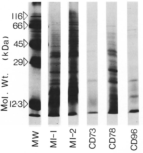
SDSPAGE of batches MI-1, MI-2, CD73, CD78,and CD96 is shown in Figure 1. MI-1 and MI-2, to which PMSF was added, have consistently resolved into comparable protein band patterns. The other batches showed variations in the overall number of bands resolved, as well as in their patterns. For example, CD96 showed a loss ofbands in the molecular weight range of 22 kDa to 65 kDa. CD73 (prepared by a different purification protocol (12) and used as a skintest reagent) resolved into only four bands of molecular weights 36, 28, 20, and 10 kDa. CD78, CD91, and CD99 were consistently comparable to each other and to MI-1 and MI-2; whereas CD84 showed a loss ofbands ofmolecular weight more than 40 kDa (data not shown).

Fig. 1. SDS-PAGE analysis of cell-free extract of M. leprae; 20 μg of protein was loaded into each well of the gel.
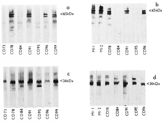
Four IgG monoclonal antibodies (supplied by the WHOIMMLEP bank) were used to study the different batches of CFX of M. leprae by immunoblotting. Y1-2 and IIIE9 (5) recognize different epitopes on the 65-kDa molecule. IIIE9 is M. leprae specific; whereas Y1-2 is crossreactive with other mycobacteria. The 65kDa band was readily detectable in MI-1, MI-2, CD78, CD91, CD96, and CD99; whereas batches CD73, CD84, and CD95 lacked such a band (The Table; Fig. 2a and b). A band of 40kDa molecular weight was detected in CD95 by IIIE9, but not by Y1-2. Several other lower molecular weight bands were recognized, all of which were less intense and slower to develop color than the major bands of 65 kDa and 40 kDa. Monoclonal antibody F479 (5) recognizes an epitope on the 36kDa protein. All of the batches tested reacted with this antibody (The Table; Fig. 2c and d). However CD96 and CD73 showed faint bands compared to the others, and CD95 showed a lower molecular weight band in addition to the 36kDa band. The monoclonal antibody SA1D2D1 (13) recognizes an epitope on the 28-kDa protein in addition to some higher molecular weight bands. All of the batches of CFX reacted with this antibody, although CD84 and CD96 showed fainter bands than the other preparations.

Fig. 2. Immunoblot analysis of different batches of cell-free extract of M. leprae using monoclonal antibodies Y1-2 (a and b) and F47-9 (c and d). Positions of the 65-kDa protein (a and b) and the 36-kDa protein (c and d) are shown on the right of the figure.
DISCUSSION
These data suggest that differences exist among the different batches of CFX of M. leprae. It seems unlikely that these differences result from differences in the length of time of storage, since batch CD78 was stored for the longest period of time yet contained all the major bands. Batch CD95 was prepared 6 months after CD78 but did not contain any detectable 65-kDa band. The 65-kDa protein appears to be particularly susceptible to protease activity, being stabilized by the presence of PMSF. Since PMSF inhibits serine proteases only, it is possible that, even in the presence of PMSF, other proteases will still be active. Similar, though less obvious differences were found between batches when comparisons of the 36-kDa and 28-kDa proteins were analyzed. It should be stressed that in this study we have looked only at three well-characterized M. leprae antigens; clearly a more exhaustive analysis is likely to reveal other major variations. This is supported by the findings revealed by SDS-PAGE analysis (Fig. 1).
Batches of CFX of M. leprae are being used in several laboratories throughout the world as a source of M. leprae antigens. We report these data to emphasize the variability which may exist between different batches of this material, and hence the need for caution when comparing data from different laboratories.
Acknowledgments. M.I. was supported by the British Leprosy Relief Association (LEPRA). We wish to thank Dr. R. J. W. Rees and Mrs. Celia Lowe for providing many of the bacterial extracts, and for their helpful comments throughout the study.
REFERENCES
1. BRITTON, W. J., HELLQVIST, L., BASTEN, A. and RAISON, R. L. Mycobacterium leprae antigens involved in human immune responses. 1. Identification of four antigens by monoclonal antibodies. J. Immunol. 135(1985)4171-4177.
2. CHAKRABARTY, A. K., MAIRE, M. A. and LAMBERT, P. H. SDS-PAGE analysis of Mycobacterium leprae protein antigens reacting with antibodies from sera from lepromatous patients and infected armadillos. Clin. Exp. Immunol. 47(1982)523-531.
3. DRAPER, P. Protocol 1/79 purification of M. leprae. In: Report of the Fifth Meeting of the Scientific Working Group on the Immunology of Leprosy (IMMLEP). Geneva: World Health Organization, 1980, Annex 4, p. 23. TDR/IMMLEP-SWG (5)/80.3.
4. EHRENBERG, J. P. and GEBRE. N. Analysis of the antigenic profile of Mycobacterium leprae: cross-reactive and unique specificity of human and rabbit antibodies. Scand. J. Immunol. 26(1987)673-681.
5. ENGERS, H. D., ABE, M., BLOOM, B. R., MEHRA, V., BRITTON, W., BUCHANAN, T. M., KHANOLKAR, S. R., YOUNG, D. B., CLOSS, O., GILLIS, T., HARBOE, M., IVANYI, J., KOLK, A. H. J. and SHEPARD, C. C. Results of a World Health Organisation-sponsored workshop on monoclonal antibodies to Mycobacterium leprae. (Letter) Infect. Immun. 48(1985)603-605.
6. KIRCHHEIMER, W. F. and STORRS, E. E. Attempts to establish the armadillo (Dasypus novemcinctus Linn.) as a model for the study of leprosy. 1. Report of lepromatoid leprosy in an experimentally infected armadillo. Int. J. Lepr. 39(1971)693-702.
7. KLATSER, P. R., VAN RENS, M. and EGGELTE, T. A. Immunochemical characterisation of Mycobacterium leprae antigens by the SDS-PAGE immunoperoxidase technique (SGIP) using patients' sera. Clin. Exp. Immunol. 56(1984)537-544 .
8. KYHSE-ANDERSEN, J. Electroblotting of multiple gels: a simple apparatus without buffer tank for rapid transfer of proteins from polyacrylamide to nitrocellulose. J. Biochem. Biophys. Methods 10(1984)203-209.
9. LAEMMLI, U. K. Cleavage of structural proteins during the assembly of the head of bacteriophage T4. Nature (London) 227(1970)680-685.
10. LEVIS, W. R., MEEKER, H. C, SCHULLER-LEVIS, G. B., GILLIS, T. P., MARINO, L. J., JR. and ZABRISKIE, J. Serodiagnosis of leprosy: relationships between antibodies to Mycobacterium leprae phenolic glycolipid I and protein antigens. J. Clin. Microbiol. 24(1986)917-921.
11. LOWRY, O. H., ROSEBROUGH, N. J., FARR, A. L. and RANDALL, R. J. Protein measurement with the Folin-phenol reagent. J. Biol. Chem. 193(1951)265-275.
12. SHEPARD, C. C. DRAPER, P., REES, R. J. W. and LOWE, C. Effect of purification steps on the immunogenicity of Mycobacterium leprae. Br. J. Exp. Pathol. 61(1980)376-379 .
13. YOUNG, D. B., FOHN, M. J., KHANOLKAR, S. R. and BUCHANAN, T. M. Monoclonal antibodies to a 28,000 mol. wt. protein antigen of Mycobacterium leprae. Clin. Exp. Immunol. 60(1985)546-552.
1. M.B., B.Ch., M.Sc, Head, Laboratory for Leprosy and Mycobacterial Research, National Institute for Medical Research, The Ridgeway, Mill Hill, London NW7 1AA, U.K. Present address for Dr. Ibrahim is Bland Sutton Institute of Pathology, University College and Middlesex School of Medicine, London, U.K.
2. B.Sc, Ph.D., Head, Laboratory for Leprosy and Mycobacterial Research, National Institute for Medical Research, The Ridgeway, Mill Hill, London NW7 1AA, U.K. Present address for Dr. Ibrahim is Bland Sutton Institute of Pathology, University College and Middlesex School of Medicine, London, U.K.
3. B.Sc, Ph.D., Head, Laboratory for Leprosy and Mycobacterial Research, National Institute for Medical Research, The Ridgeway, Mill Hill, London NW7 1AA, U.K. Present address for Dr. Ibrahim is Bland Sutton Institute of Pathology, University College and Middlesex School of Medicine, London, U.K.
Reprint requests to Dr. Colston.
Received for publication on 10 May 1988.
Accepted for publication in revised form on 24 October 1989.