- Volume 57 , Number 1
- Page: 1–11
HLA-DR-restricted antigen-induced proliferation and cytotoxicity mediated by CD4+ T-cell clones f rom subjects vaccinated with killed M. leprae
ABSTRACT
Thirteen CD4+ T-cell clones raised against Mycobacterium leprae f rom three M. leprae-vaccinated subjects were studied for major histocompatibility complex (MHC) restriction in proliferative and cytotoxicity assays. These T-cell clones recognized at least nine different epitopes, ranging f rom M. leprae-specific to broadly crossreactive. Restriction studies with a panel of antigen-presenting cells (APCs) suggest that all of the T-cell clones recognized antigens in the context of the DR locus. Three T-cell clones with three different reactivities f rom a DR1, 2-positive subject responded to M. leprae in proliferation and cytotoxicity when the antigen was presented in the context of DR1-positive APCs. Four T-cell clones responding to M.leprae-specific or crossreactive epitopes f rom the second donor, who was DR4.DW4; DR4,Dw14-positive, and a single M. leprae-specific T-cell clone f rom the third subject, who was DR3,4:Dw4, recognized the antigens in the presence of Dw4 APCs. Four crossreactive T-cell clones f rom the second subject responded in the presence of Dw14-positive APCs, and one limited crossreactive clone recognized the antigen in the context of DR4 and DR7 positive cells, suggesting that its response was restricted by a common determinant. The T-cell clones that recognize the 65-kDa, 18-kDa, and 13B3 recombinant M. leprae antigens in proliferative assays were cytotoxic for autologous adherent cells pulsed with the respective antigens. The response of the respective T-cell clones to the recombinant antigens in proliferation and cytotoxicity was restricted by similar class II molecules as for killed M. leprae, i.e., the 65-kDa antigen was recognized in the context of DR 1, the 18-kDa antigen in the context of DR4,Dw4, and the 13B3 antigen in the context of DR4 and DR7 class II MHC molecules. The experiments with Dw4 and Dw14-positive mixed adherent cells and appropriate T-cell clones suggest that cytotoxicity requires direct interaction between CD4+ T cells and antigen-presenting cells.RÉSUMÉ
On a étudié 13 clones de cellules T CD4+ dirigés contre Mycobacterium leprae, provenant de trois sujets vaccinés contre M. leprae, en ce qui concerne la restriction du complexe majeur d'histocompatibilité (MHC), dans des essais de prolifération et de cytotoxicité. Ces clones de cellules T reconnaissaient au moins neuf épitopes difiêrents, qui allaient d'épitopes spécifiques pour M. leprae, à des épitopes témoignant d'une réactivité croisée étendue. Des études de restriction, menées avec une batterie de cellules qui présentent les antigènes (APCs), suggèrent que tous les clones de cellules T reconnaissent les antigènes dans le contexte du locus DR. Trois clones de cellules T ayant trois profils de réactivité différents, et provenant d'un sujet DR1, 2-positif, répondaient à M. leprae lors des essais de prolifération et de cytotoxicité, lorsque l'antigène était présenté dans le contexte de cellules présentant les antigènes qui étaient DR 1-positives. Quatre clones de cellules T réagissant à des épitopes spécifiques pour M. leprae ou de réactivité croisée, en provenance du second donneur, qui était DR4, Dw4, DR4, Dw14-positif, et un seul clone de cellules T spécifiques pour M. leprae en provenance du troisième sujet, qui était DR3,4, Dw4, reconnurent les antigènes en présence de cellules APC Dw4. Quatre clones de cellules T avec réactivité croisée, provenant du deuxième sujet, répondaient en présence de cellules APC Dw14 positives, et un clone à réactivité croisée limitée, reconnaissait l'antigène dans le contexte de cellules positives DR4 ou DR7, ce qui suggère que leur réponse était contrôlée par un déterminant commun. Les clones de cellules T qui reconnaissaient les antigènes recombinants 65-kDa, 18-kDa, et 13B3 de M. leprae au cours d'essais de prolifération, étaient cytotoxiques pour des cellules adhérentes autologues stimulées par les antigènes respectifs. La réponse des clones respectifs de cellules T aux antigènes recombinants, lors des essais de prolifération et de cytotoxicité, était contrôlée par des molécules du complexe majeur d'histocompatiabilité de classe II semblables à celles qui sont impliquées dans la réaction à M. leprae tué, c'est-à-dire que l'antigène 65-kDa était reconnu dans le contexte DR 1, l'antigène 18-kDa dans le contexte DR4, Dw4, et l'antigène 13B3 dans le contexte des molécules MHC de classe II DR4 et DR7. Les expériences menées avec un mélange de cellules adhérentes positives pour Dw4 et Dw14, et des clones de cellules T correspondants, suggèrent que la cytotoxicité requiert une interaction directe entre les cellules T CD4+ et les cellules qui présentent l'antigène.RESUMEN
Utilizando ensayos de proliferación y citotoxicidad, se estudió la restricción por el complejo mayor de histocompatibilidad (CMH), en trece clonas de células T CD4+ generadas contra Mycobacterium leprae en tres sujetos vacunados con M. leprae. Estas clonas de células T reconocen cuando menos 9 epítopes diferentes, algunos específicos para M. leprae, otros con reacción cruzada. Los estudios de restricción con un panel de células presentadoras de antigenos (CPA) sugieren que todas las clonas de células T reconocen antígenos en el contexto del locus DR. Tres clonas de células T, con tres diferentes reactividades, de un sujeto DR1, 2-positivo, respondieron a M. leprae en los ensayos de proliferación y citotoxicidad cuando el antígeno fue presentado por CPA DR 1-positivas. Cuatro clonas de células T respondieron a epitopes específicos de M. leprae o a epítopes de reacción cruzada del segundo donador, quien fue DR4, Dw4; DR4. Dw14-positivo, y una sola clona específica para M. leprae un tercer sujeto, quien fue DR3, 4, Dw4, reconoció los antígenos en presencia de CPA Dw4. Cuatro clonas de células T de reacción cruzada del segundo sujeto respondieron en presencia de CPA Dw14-positivas, y una clona de reactividad cruzada limitada reconoció al antígeno en células DR4- y DR7-positivas, sugiriendo que su respuesta fue restringida por un determinante común. Las clonas de células T que reconocen a los antígenos recombinantes 65-kDa, 18-kDa y 13B3, de M. leprae en los ensayos de proliferación, fueron citotoxicos para células adhérentes autólogas estimuladas con los antígenos respectivos. Al igual que la respuesta contra M. leprae muerto, la respuesta de las clonas de células T hacia los antígenos recombinantes en los ensayos de proliferación y citotoxicidad, estuvo restringida por moléculas CMH clase II similares, esto es, el antígeno 65-kDa fue reconocido en el contexto de DR1, el 18kDa en el contexto de DR4, D\v4, y el 13B3 en el contexto de DR4 y DR7. Los experimentos con células adhérentes mezcladas Dvv4 y Dw14 y las clonas de células T apropiadas, sugieren que la citotoxicidad requiere de la interacción directa entre las células T CD4 + y las células presentadoras de antígeno.CD4+ T cells upon interaction with specific antigens produce biologically important lymphokines required for activation and proliferation of cells responsible for cell-mediated immune (CMI) responses (4,15,16,24) The antigens recognized by CD4+ T cells may have potentials as vaccines for diseases like leprosy where a strong correlation exists between CMI and resistance against the disease. Isolation of the genes encoding for major Mycobacterium leprae protein antigens (33) has led to the identification of recombinant M. leprae proteins recognized by M. leprae-reactive CD4+ T-cell clones in proliferative assays (17, 20, 21, 9, 26). Such recombinant antigens as vaccines may have an advantage over whole, killed M. leprae which have epitope(s) capable of activating suppressor-T cells (14, 18). However, CD4+ T cells recognize antigens in the context of highly polymorphic class II major histocompatibility complex (MHC) molecules (10). As a first step toward understanding MHC restriction in antigen recognition by CD4+ T-cell clones established from the volunteers vaccinated with killed M. leprae, we have studied their responses against a panel of antigen-presenting cells.
Like viral infections, cytotoxicity mediated by the immune cytotoxic T cells against mycobacteria-infected targets may be one of the mechanisms involved in protection (8). We have demonstrated the cytotoxic function off M. leprae-induced CD4 + T-cell clones established from killed-M. leprae-vaccinated subjects (20, 26). From among the CD4+ T-cell clones mediating cytotoxicity there were also T-cell clones recognizing the recombinant antigens in the proliferative assays (20, 26). However, it was not known whether the same or different antigens were required for proliferative and cytotoxic activities and if single or multiple class II MHC molecules were the restricting elements. Here we report that a given recombinant antigen mediates both functions, and that recognition of the antigens for proliferative and cytotoxic activities is restricted by HLA-DR molecules.
MATERIALS AND METHODS
T-cell clones. Forty-two CD4+ T-cell clones were established from the peripheral blood of human volunteers vaccinated with killed M. leprae (21) Thirteen of these T-cell clones from three different subjects were used in the present study. The T-cell clones A7, 3C7 and A8 were established from donor JM. The T-cell clones 2/3D, 4/1F, 4/4C, 5/2B, 5/6B, 1/6B, 6/9B, 6/10F and 2/4F were raised from donor ATT. The T-cell clone 2/2F was established from donor NHH and has a reactivity pattern similar to the ATT clones 6/1 OF and 2/4F (21). From among the six major recombinant M. leprae antigens identified by antibody probes (33), the 65-kilodalton (kDa) antigen induced proliferation of the T-cell clone JM A7 (26), the 18-kDa antigen induced proliferation of the T-cell clones ATT 6/1 OF, 2/4F and NHH 2/2F(21). The ATT T-cell clone 2/3D responded to a recombinant antigen 13B3 identified by use of human T-cell clones as primary probes to isolate recombinant antigens (25).
Antigens. Killed M. leprae was kindly provided by Dr. R. J. W. Rees, Clinical Research Centre, Harrow, England, from the WHO/IMMLEP bank. All other mycobacteria were kindly supplied by Dr. Otto Closs, National Institute of Public Health, Oslo, Norway. The bacilli were killed by irradiation. M. leprae was used at.5 x 107 bacilli/ml and all other mycobacteria were used at 10 µg/ml. Escherichia coli lysates containing the 65-kDa, 18-kDa and 13B3 antigen were prepared according to protocols described earlier (21, 27). The lysates at a protein concentration of 50 µg/ml were used for proliferative and cytotoxicity assays.
Antigen-presenting cells (APCs). Adherent cells from autologous and allogeneic peripheral blood mononuclear cells (PBMC) from a panel of donors were used as antigen presenting cells (APCs) for proliferative and cytotoxicity assays. All APCs were typed for HLA-A, 13, C and DR, and DR4 cells were typed for Dw specificities as described by Qvigstad, et al. (30).
Proliferative assay. Samples of 1 x 10⁴ cells of individual T-cell clones were added to the wells of 96-well plates (Costar, Cambridge, Massachusetts. U.S.A.) and stimulated with an optimal concentration of antigen in the presence of adherent cells from 1 x 10⁵ irradiated PBMC as described earlier (24). The plates were incubated at 37ºC in a humidified atmosphere of 5% CO, and 95% air, and 0.045 MBq 3H-thymidine was added to the cultures during the last 4 hr of a 72-hr incubation period. Cultures were harvested on a Skatron automatic harvestor, and the radioactivity incorporated was determined by standard methods (22). Median counts per minute (cpm) values have been used to express the results. A clone was considered responding to a given antigen when Δcpm was > 1000 and T/C >2. Such values are underlined in the tables. Δcpm and T/C are defined as:

and

Cytotoxicity assay. A neutral red uptake assay, as described earlier (23, 26) was used to assess the cytotoxic potential of T-cell clones against antigen-pulsed autologous and allogeneic adherent cells. In brief, adherent cells from 1 x 10⁶ irradiated PBMC in the individual wells of 24-well Costar plates were pulsed with killed M. leprae or recombinant antigens, and 1 x 10⁵ cloned T cells were added to each well. The plates were incubated for 7 days at 37ºC in a humidified atmosphere of 5% CO2 and 95% air. At the end of the incubation period, nonadherent cells were washed off. The remaining macrophages differentiated from adherent monocytes were allowed to take up neutral red. The dye taken up by the macrophages was released in a 0.5 ml solution of 0.05 M acetic acid in 50% ethanol, and was quantitated by measuring OD 540 in a spectrophotometer. Results are expressed in terms of percentage cytotoxicity which is defined as:

where the OD 540 control = OD 540 of cultures with adherent cells + antigen, and the OD 540 experimental = OD 540 of cultures with adherent cells + T-cell clone + antigen.
RESULTS
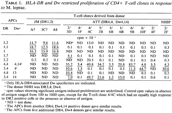
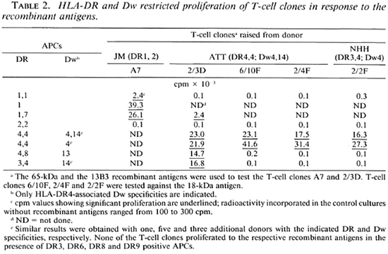
All 13 T-cell clones were tested with a panel of mycobacteria using autologous APCs in proliferative assays to have an idea of the number of possible epitopes recognized by them. The T-cell clones from donor JM recognized three different epitopes. The T-cell clones from donors ATT and NHH recognized at least six different epitopes (data not shown). To determine the MHC restriction of antigen recognition, all T-cell clones were tested for M. leprae-induced proliferation against autologous and a panel of allogeneic APCs (Table 1). Since the T-cell clones recognized the antigens in the context of DR molecules or Dw specificities of DR4, only these phenotypes of the APCs are tabulated. The three T-cell clones from donor JM (who is DR1,2) recognized antigens in the context of DR 1 (Table 1). The T-cell clones from donor ATT (who is DR4 Dw4; DR4 Dw14) showed three different restriction patterns. The T-cell clone 2/3D recognized antigen in the presence of APCs that express serologically defined DR4 and DR7 molecules (Table 1). Other T-cell clones responded in the presence of either DR4, Dw4 or DR4, Dw14 positive APCs. The T-cell clones 4/1F, 6/9B, 6/1 OF and 2/4F, which recognized at least three different epitopes of M. leprae, proliferated to M. leprae in the context of DR4, Dw4 and the remaining ATT T-cell clones- 4/4C, 5/2B, 5/6B and 1/6B that recognized at least two different epitopes-responded to M. leprae antigens in the presence of DR4, Dw14 positive APCs (Table 1). In addition, the T-cell clone 4/4C proliferated to DR2 positive cells in the absence of M. leprae antigens. The T-cell clone 2/2F from donor NHH (who is DR3,4; Dw4) recognized M. leprae antigen in the presence of DR4, Dw4 positive APCs (Table 1). An analysis of the proliferative responses of these T-cell clones to relevant recombinant antigens showed that the T-cell clone A7 recognized the 65kDa recombinant M. leprae antigen in the presence of DR1 positive APCs. The T-cell clones 6/1 OF, 2/4F and 2/2F recognized the 18-kDa recombinant M. leprae antigen in the presence of DR4, Dw4 positive APCs and the T-cell clone 2/3D recognized the 13B3 antigen when presented by DR4 and DR7 positive APCs (Table 2). Thus, restriction elements for the recognition of relevant recombinant antigens by the T-cell clones were similar to those for the recognition of killed M. leprae antigens.
CD4+ T-cell clones that proliferate in response to M. leprae are cytotoxic for M. leprae-pulsed autologous adherent cells (20,26). Among these are the T-cell clones A7; 2/4F, 6/1 OF and 2/3D which proliferate to the 65-kDa, 18-kDa and 13B3 recombinant antigens, respectively. To determine the antigens required for cytotoxicity, autologous APCs were pulsed with E. coli lysates lacking or containing the recombinant antigens. None of these T-cell clones could significantly kill the adherent cells pulsed with the E. coli lysate lacking recombinant antigens (Table 3). The T-cell clones A7; 2/4F, 6/1 OF and 2/3D killed adherent cells pulsed with the lysatcs containing 65-kDa, 18-kDa and 13B3 recombinant antigens, respectively. These results confirm that a single antigen is recognized by the T-cell clones in both proliferative and cytotoxic activities.
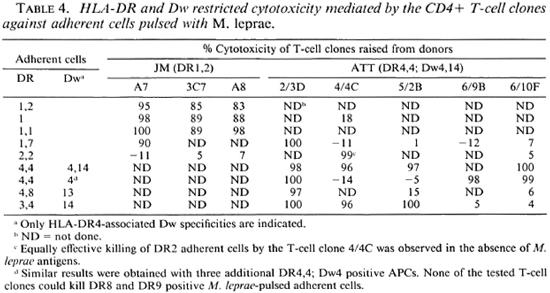
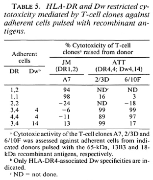
MHC restriction of cytotoxicity was determined by assessing the cytotoxic activity of the CD4+ T-cell clones against M. leprae-pulsed adherent cells from a panel of allogeneic donors. The T-cell clones A7, 3C7 and A8 killed M. leprae-pulsed adherent cells in the context of DR 1. The T-cell clone 2/3D killed DR4 and DR7 positive M. leprae pulsed adherent cells. The T-cell clones 6/9B and 6/1 OF killed M. leprae-pulsed DR4, Dw4 adherent cells. The T-cell clones 4/4C, 5/2B and 5/6B killed M. leprae adherent cells in the context of the DR4, Dw14 molecule (Table 4). In addition, the T-cell clone 4/4C showed antigen nonspecific killing of DR2 adherent cells (Table 4). Experiments with the recombinant antigens showed that killing of the APCs in the presence of the 65-kDa antigen by T-cell clone A7 was DR1 restricted. The T-cell clone 6/10F in the presence of the 18-kDa antigen killed DR4, Dw4 positive APCs, and killing by the T-cell clone 2/3 D in the presence of the 13B3 antigen was DR4 restricted and did not depend upon the Dw phenotype (Table 5).
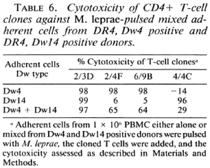
To determine the possible mechanism(s) of cytotoxicity, M. leprae-pulsed mixed adherent cells from Dw4 and Dw14 positive donors were used as targets for the T-cell clones that killed cither both or only one of these targets, when tested individually. Cells of the T-cell clone 2/3D, which killed individual adherent cells of both phenotypes, were equally effective in killing mixed adherent cells. The T-cell clones 4/4C, 6/9B and 2/4F, which were selective in killing individual adherent cells, were partially effective in killing mixed adherent cells (Table 6). Similar results were obtained with use of the recombinant antigens (data not shown).
DISCUSSION
In this study we used 13 M. leprae-responding CD4+ T-cell clones from three healthy subjects vaccinated with killed M. leprae. Their reactivity patterns with a panel of mycobacteria in proliferative assays suggest that these T-cell clones recognized at least nine different epitopes. Included in these are the T-cell clones that respond to the65-kDa, 18-kDaand 13B3 recombinant M. leprae antigens. The 65-kDa antigen represents highly crossreactive antigens (26). The 18-kDa antigen is a representative of M. leprae-specific antigens (21), and the 13B3 antigen is an example of antigens which have limited crossreactivity with other mycobacteria (25). The exhibition of cytotoxic activity by all of these T-cell clones suggests that cytotoxicity mediated by CD4+ T-cell clones is not against some specific antigens but is a general characteristic of M. leprae reactive CD4+ T cells. In our earlier studies using different species of mycobacteria, M. leprae- induced CD4+ T-cell clones exhibited an identical pattern of antigen recognition in proliferation and cytotoxic functions (20, 26). Our present study demonstrates that the T-cell clones which proliferate to the 65-kDa, 18-kDa or 13B3 recombinant antigens kill adherent cells pulsed with the respective antigens. Thus, the use of recombinant antigens confirms that a single antigen is responsible for both functions. Although killing of antigen-pulsed APCs by mycobacteria-induccd CD4+ T-cell clones has been earlier described by us and others (13, 16, 20, 23, 26) using the recombinant antigens we have experimentally demonstrated the identity of some of the antigens that mediate cytotoxicity.
CD4+ T cells after recognition of antigen on the surface of APCs arc activated to produce lymphokines like interleukin-2 (IL-2) and interferon gamma (interferon-γ). IL-2 is required for the expansion and enhancement of cytolytic activity of CD8+ T cells, which are considered to play a key role in protection against viral (10,34) and parasitic infections (32). Interferon-γ is a macrophage activator, and activated macrophages have the enhanced ability to destroy ingested intracellular parasites (8, 15, 16). This could be one mechanism by which CD4 + T cells may have a role in protection against a disease such as leprosy. The cytotoxic activity of CD4 + T cells would suggest that, like CD8 + cytotoxic T cells, these cells may also have a direct role in protection by killing infected macrophages and thereby inhibiting intracellular bacillary growth (8).
CD4+ T cells recognize antigens in the context of class II MHC molecules (10). Although there exist at least three different families of functional class II MHC molecules (namely, DP, DQ and DR), recognition of M. leprae antigens in proliferative assays by CD4+ T-cell clones from leprosy patients is primarily restricted by DR (29). Our studies show that DR molecules are the restriction elements for the proliferative and cytotoxic activities of the CD4+ T-cell clones raised from M. leprae-vaccinated healthy subjects. DR-restricted recognition of M. leprae antigens is not limited to CD4 + T cells. Modlin, et al. (19) have reported DR4-restricted suppressor activity of CD8 + suppressor-T cells isolated from lepromatous leprosy patients.
Most interesting results were obtained with the T-cell clones from the DR4 positive donor ATT. The DR4 haplotype is complex and at least five distinct specificities-Dw4, DwlO, Dw13, Dw14 and Dwl5 -have been defined (7). The T-cell clones from the Dw4, Dw14 positive donor ATT showed three different restriction patterns. Antigen 13B3 was recognized by the T-cell clone 2/3D in the context of serologically defined DR4. The response of other T-cell clones was restricted by either Dw4 or Dw14 positive APCs. Dw4-restricted T-cell clones include the T-cell clones that respond to the 18-kDa M. leprae protein and two other epitopes whose identification is unknown. Dw14- specific T-cell clones recognized at least two different hitherto unknown antigens/epitopes. In another study with human CD4+ T-cell clones from a leprosy patient who was DR3,4; Dw13, Ottenhoff, et al. (24) have shown that one of the six M. leprae-reactive T-cell clones recognized the antigen in association with a restriction determinant that was closely associated with DR4 and not related to the Dw specificity of the APCs; whereas the other five were restricted by determinants associated with Dw specificity, namely, Dw13.
Serologically defined, DR4 is associated with the αβ dimer. The α chain is nonpolymorphic. Amino-acid sequences deduced from the nucleotide sequence of DRβ1 cDNA clones show that the cells with Dw4, Dw13 and Dw14 specificities differ from each other in only one to three amino acids at positions 71, 74, and 86. Dw4 and Dw 13 differ at all of these three positions. Dw4 and Dw14 have different amino acids at positions 71 and 86. Dw13 and Dw14 differ in only one amino acid at position 74 (7). Specificity of the response of M. leprae-reactive CD4+ T-cell clones in the context of different Dw specificities of DR4 would suggest that processed antigenic peptides of M. leprae can distinguish even a difference of one amino acid for binding with MHC molecules and, therefore, all three of the DR4 specificities studied (Dw4, Dw 13 and Dw 14) are important for the recognition of different M. leprae antigens. Because the β 1 chain from these specificities differs in only one to three amino acids between positions 71 and 86, this may be the region where processed peptides bind for recognition by the T cells. The T-cell clones that recognize M. leprae antigens in the context of Dw4, Dw 14 and Dw13 showed at least three, two and five different reactivity patterns, respectively, suggesting that it was not just one antigenic peptide that could differentiate between the above-discussed amino-acid substitutions but several, probably as many as the different reactivities possessed by the T-cell clones.
The T-cell clone 4/4C from donor ATT, which showed an antigen-specific response in the presence of DR4, Dw14 positive APCs, responded in the proliferative and cytotoxicity assays to DR2 positive APCs in the absence of specific antigen. In a recent study, a T-cell clone recognizing an M. leprae peptide in the context of the DR2 molecule responded to a structurally similar synthetic peptide from the DR2 molecule itself (1). A similar mechanism may be responsible for the DR2 alloreactivity of the T-cell clone 4/4C.
Our experiments with the mixed adherent cells from Dw4 and Dw14 positive donors showed that the T-cell clone 2/3D, which killed either of the adherent cells when tested individually, was equally effective in killing the mixed adherent cells. The other T-cell clones that killed either Dw4 or Dw14 positive adherent cells were only partially effective in killing the mixed adherent cells (Table 6). These results suggest that the cytotoxicity mediated by CD4+ T cells is an active phenomenon which requires direct recognition of the target cells. These results also support our earlier findings (23) that the cytotoxic activity of the CD4+ T cells is not mediated via nonspecific factors and that the lower dye uptake in the experimental wells is not due to loss of macrophage adherence after activation with lymphokines or mycobacterial antigens.
The development of an effective antileprosy vaccine is necessary for the control and eradication of leprosy. Killed M. leprae obtained from armadillo tissues is one of the candidate antileprosy vaccines. Sensitization studies in healthy human volunteers from a nonendemic country demonstrated that M. leprae-specific CMI responses are induced in a majority of the people vaccinated with killed M. leprae (11, 2). However, M. leprae has epitopes that can activate suppressor cells C (4, 18 ). To what extent such suppressor epitopes can down regulate or interfere withM. leprae-mduced CMI responses in individuals in leprosy-endemic areas is yet to be determined. Other potential limitations may be the cost and supply of sufficient quantities of armadillo-derived M. leprae. The identification of recombinant M. leprae antigens by CD4+ T-cell clones has generated an interest in exploring the possibility of using these antigens to develop the next generation of vaccines. The precise epitopes recognized by CD4+ T cells can be mapped using synthetic peptides (28). These antigens or peptides can be expressed in suitable vectors used as a vaccine vehicle(3). Since the antigen recognition by CD4 + T-cell clones is restricted by products of the highly polymorphic class II MHC, an isolated antigen or a single peptide may be able to associate with only a limited number of MHC types ( 2, 5, 6) and invigorate the CMI responses only in a minority. On the other hand, killed M. leprae, because it has multiple antigens/epitopes that may associate with all possible MHC types, may be able to activate CMI responses in the majority of the people. However, since many specificities of M. leprae could be recognized by a single MHC molecule, all of the T-cellreactive epitopes of M. leprae capable of binding to that particular MHC may not be necessary to induce M. leprae responsiveness. The identification of more antigens like 13B3, which can activate T cells in the context of more than one DR type and to subtypes of complex haplotypes, in this case DR4, is necessary. The use of a mixture of such peptides may be able to induce CMI responses in recipients with varied MHC backgrounds (31).
Acknowledgments. The material and intellectual help provided by Dr. Tore Godal, Prof. Erik Thorsby, Dr. R. J. W. Recs, Dr. Otto Closs, and Dr. Richard Young is gratefully acknowledged. Recombinant human interleukin-2 was kindly supplied by Cetus Corporation. This work was supported by grants from the UNDP/World Bank/WHO Special Programme for Research and Training in Tropical Diseases, from the Rockefeller Foundation Program for Research in Great Neglected Diseases, and the Laurine Maarschalk Fund.
REFERENCES
1. Anderson, D. C, van Schooten, W. C. A., Barry, M. E., Janson, A. A. M., Buchanan, T.M. and de Vries, R. R. P. A Mycobacterium leprae-specific human T cell epitope cross-reactive with an HLA-DR2 peptide. Science 242(1988)259-261.
2. Babbitt, B. P., Matsueda, G., Haber, E., Unanue, E. R. and Allen, P. M. Antigenic competition at the level of peptidc-Ia binding. Proc. Natl. Acad. Sci. U.S.A. 83(1986)4509-4513.
3. Bloom, B.R. Learning from leprosy: a perspective on immunology and the Third World. J. Immunol. 137(1986)i-x.
4. Boom, W. H., Husson, R. N., Young, R. A., David, J.R. and Piessens, W.F. In vivo and in vitro characterization of murine T cell clones reactive to Mycobacterium tuberculosis. Infect. Immun. 55(1987)2223-2229.
5. Buss, S., Colon, S., Smith, C, Freed, J. H., Miles, C. and Grey, H.M. Interaction between a "processed" ovalbumin peptide and la molecule. Proc. Natl. Acad. Sci. U.S.A. 83(1986)3968-3971.
6. Buss, S., Sette, A., Colon, S.M., Miles, C. and Grey , H. M. The relation between major histocompatibility complex (MHC) restriction and the capacity of la to bind immunogenic peptides. Science 235(1987)1353-1358.
7. Cairns, J. S., Curtsinger, J.M., Dahl, C. A., Freeman, S., Alter, B. J. and Bach, F.H. Sequence polymorphism of HLA DRβ1 alleles relating to T-cell recognized determinants. Nature 317(1985)166-168.
8. De Lihero, G., Flesch, I. and Kaufmann, S.H.E. Mycobacteria reactive Lyt-2+ T cell lines. Eur.J. Immunol.18(1988)59-66.
9. De Vries, R. R. P., Ottenhoff, T. H.M., Li, S.-G. and Young R. A. HLA class II restricted helper and suppressor clones reactive with Mycobacterium leprae. Lepr. Rev. 57 Suppl. 2(1986)113-121.
10. Fitch, R. W. T-cell clones and T-cell receptors. Microbiol. Rev. 50(1986)50-69.
11. Gill, H. K., Mustafa, A. S. and Godal, T. Induction of delayed-type hypersensitivity in human volunteers immunized with a candidate leprosy vaccine consisting of killed Mycobacterium leprae. Bull. WHO 64(1966)121-126.
12. Gill, H. K., Mustafa, A. S. and Godal, T. In vitro proliferation of lymphocytes from human volunteers vaccinated with armadillo-derived, killed M. leprae. Int. J. Lepr.55(1987)30-35.
13. Hansen, P. W., Petersen, C.M., Povlsen, J. V. and Kristensen, T. Cytotoxic human HLA class II restricted purified protein derivative reactive T lymphocyte clones.IV. Analysis of HLA restriction pattern and mycobacterial antigen specificity. Scand J.Immunol. 25(1987)295-303.
14. Kaplan, G., Gandhi, R. J., Weinstein, D. E., Levis, W. R., Patarroyo, M.E., Brennan, P.J. and Cohn, Z. A. Mycobacterium leprae antigeninduced suppression of T cell proliferation in vitro. J. Immunol. 138(1987)3028-3034.
15. Kaufmann, S. H. E. Possible role of helper and cytolytic T lymphocytes in antibacterial defense: conclusions based on a murine model of listeriosis. Rev. Infect. Dis. 9 Suppl. 5(1987)S650-S659.
16. Kaufmann, S. H. E., Chiplunkar, S., Flesch, I. and De Libero, G. Possible role of helper and cytolytic T cells in mycobacterial infections. Lepr. Rev. 57 Suppl. 2(1986)101-111.
17. Klatser, P. R., Hartskeerl, R. A;, Van Schooten, W. C. A., Kolk, A. H. J. Van Rens, M. M. and De Wit , M. Y. L . Characterization o f the 36 K antigen of Mycobacterium leprae. Lepr. Rev. 57 Suppl. 2(1986)77-81.
18. Mehra, V., Brennan, P. J., Rada, E., Convit, J. and Bloom, B. R. Lymphocyte suppression in leprosy induced by unique M. leprae glycolipid. Nature 308(1984)194-196.
19. Modlin, R. L., Kato, H., Mehra, V., Nelson, E. E., Fran, X.-D., Rea, T. H., Pattengale, P. K. and Bloom, B. R. Genetically restricted suppressor T cell clones derived from lepromatous leprosy lesions. Nature 322(1986)459-461.
20. Mustafa, A. S. Identification of T cell activating recombinant antigens shared between three candidate antileprosy vaccines, killed M. leprae, My cobacterium bovis BCG and Mycobacterium w. Int. J. Lepr. 56(1988)265-273.
21. Mustafa, A. S., Gill, H. K., Nerland, A., Britton, W. J., Mehra, V., Bloom, B. R., Young, R. A. and Godal, T. Human T cell clones recognize a major M. leprae protein antigen expressed in E. coli. Nature 319(1986)63-66.
22. Mustafa, A. S. and Godal, T. In vitro induction of human suppressor T cells by mycobacterial antigens. BCG activated OKT4+ cells mediate suppression of antigen induced T cell proliferation. Clin. Exp. Immunol. 52(1983)29-37.
23. Mustafa, A. S. and Godal, T. BCG induced CD4+ cytotoxic T cells from BCG vaccinated healthy subjects: relation between cytotoxicity and suppression in vitro. Clin. Exp. Immunol. 69(1987)255-262.
24. Mustafa, A. S., Kvalheim, G., Degre, M. and Godal, T. Mycobacterium bovis BCG induced human T-cell clones from BCG-vaccinated healthy subjects: antigen specificity and lymphokine production. Infect. Immun. 53(1986)491-497.
25. Mustafa, A. S., Oftung, F., Deggerdal, A., G>ill, H. K., Young, R. A. and Godal, T. Gene isolation with human T lymphocyte probes. Isolation of a gene that expresses an epitope recognized by T cells specific for Mycobacterium bovis BCG and pathogenic mycobacteria. J. Immunol. 141(1988)2729-2733;
26. Mustafa, A. S., Oftung, F., Gill, H. K. and Natvig, I. Characteristics of human T-cell clones from BCG and killed M. leprae vaccinated subjects and tuberculosis patients. Lepr. Rev. 57 Suppl. 2(1986)123-130.
27. Oftung, F., Mustafa, A. S., Husson, R., Young, R. A. and Godal, T. Human T cell clones recognize two abundant Mycobacterium tuberculosis protein antigens expressed in Escherchia coli. J. Immunol. 138(1987)927-931.
28. Oftung, F„ Mustafa, A. S., Shinnik, T. M., Houghten, R. A., Kvalheim, G., Degre, M., Lundin, K. E. A. and Godal, T. Epitopes of the Mycobacterium tuberculosis 65-kilodalton protein antigen as recognized by human T cells. J. Immunol. 141(1988)2749-2754.
29. Ottenhoff, T. H. M., Neuteboom, S., Elferink, D. G. and de Vries, R. R. P. Molecular localization and polymorphism of HLA class II restriction determinants defined by Mycobacterium lepraereactive helper T cell clones from leprosy patients. J. Exp. Med. 164(1986)1923-1939.
30. Qvigstad, E., Thorsby, E., Reinsmoen, N. L. and Bach, F. H. Close association between the Dw14 (LD 40) subtype of HLA-DR4 and a restriction element for antigen-specific T-cell clones. Immunogenetics 20(1984)583-588.
31. Rothbard, J. Synthetic peptides as vaccines. Nature 330(1987)106-107.
32. Weiss, W. R., Sedegah, M., Beaudoin, R. L., Miller, L. H. and Good, M. F. CD8+ T cells (cytotoxic/suppressors) arc required for protection in mice immunized with malaria sporozoites. Proc. Natl. Acad. Sci. U.S.A. 85(1988)573-576.
33. Young, R. A., Mehra, V., Sweetser, D., Buchanan, T., Clark-Curtiss, J., Davis, R. W. and Bloom, B. R. Genes for the major protein antigens of the leprosy parasite Mycobacterium leprae. Nature 316(1985)450-452.
34. Zinkernagel, R. M. and Doherty, P. C. MHC restricted cytotoxic T cells: studies on the biological role of polymorphic major transplantation antigens determining T cell restriction specificity, function and responsiveness. Adv. Immunol. 27(1979)51-77.
1. Ph.D., Laboratory for Immunology, Institute for Cancer Research, The Norwegian Radium Hospital, Montebello, N-0313 Oslo 3, Norway.
2. M.D., Ph.D., Institute of Transplantation Immunology, The National Hospital, 0227 Oslo I, Norway.
Reprint requests to Dr. Mustafa at his current address: Whitehead Institute for Biomedical Research, Nine Cambridge Center, Cambridge, Massachusetts 02142, U.S.A.
Received for publication on 7 October 1988.
Accepted for publication in revised form on 9 December 1988.
