- Volume 57 , Number 1
- Page: 65–72
Light- and electron-microscopic study of M. leprae-infected armadillo nerves
ABSTRACT
Lesions in peripheral nerves of armadillos experimentally infected with Mycobacterium leprae were studied by light- and electron-microscopy. Bacilli could be found clearly inside axons of unmyelinated nerve fibers. Heavily bacillated Schwann cells were seen embracing unmyelinated axons with interrupted cytoplasmic membranes. This indicated the initiation of rupture of those cells which were responsible for the liberation of bacilli into the axons. The nerve lesions were divided into three grades according to their severity: grade I showed lesions focalized in the perineurium; grade II lesions were scattered inside nerve tissue; and in grade III lesions the nerve tissues were diffusely affected. No regressive changes, such as fibrosis or scar formation, were seen in the nerve lesions. Bacillated macrophages were not as foamy as those of human lesions, indicating that these bacillated cells were younger or more easily disrupted with a higher turnover than the cells in human lesions. This would promote the spread of lesions in armadillos, and would explain the less foamy appearance of the cells. We found bacilli inside lymphatics surrounding the nerves, substantiating the opinion that lesions spread to peripheral nerves not only by a hematogenous route but also by the lymphatics.RÉSUMÉ
On a étudié par microscopic optique et par microscopie électronique les lésions des nerfs périphériques chez des tatous infectés expérimentalement par Mycobacterium leprae. Des bacilles pouvaient être clairement mis en évidence à l'intérieur des axones des libres nerveuses non myélinisées. On a également observé des cellules de Schwann fort chargéesen bacilles, qui enserraient des axones non myélinisées tout en entraînant une interruption des membranes cytoplasmiques. Ces observations traduisent le début de la rupture des cellules responsables pour la libération des bacilles à l'intérieur des axones. Les lésions nerveuses ont été classées en trois catégories d'après leur gravité: le degré I comportait des lésions en foyers dans le périnevre; les lésions de deuxième degré étaient disséminées à l'intérieur du tissus nerveux; dans les lésions de degré III les tissus nerveux étaient atteints de manière diffuse. Aucune modification de régression telle qu'une fibrose ou la formation de cicatrices n'a été constatée dans les lésions nerveuses. Les macrophages chargésenbacilles n'étaient pas aussi spumeux que ceux que l'on observe dans les lésions chez l'homme, ce qui montre que les cellules porteuses de bacilles étaient plus jeunes, ou plus aisément bouleversées, et qu'elles étaient remplacées plus rapidement que les cellules présentes dans les lésions humaines. Ce mécanisme pourrait favoriser la dissémination des lésions chez les tatous; et pourrait également expliquer l'apparence moins spumeuse des cellules. Des bacilles ont été observés à l'intérieur des lymphatiques qui entourent les nerfs, ce qui supporte l'opinion que les lésions se propagent aux nerfs périphériques, non seulement par voie hématogène, mais aussi par les lymphatiques.RESUMEN
Se estudiaron las lesiones de los nervios periféricos de armadillos infectados experimentalmente con Mycobacterium leprae utilizando microscopía de luz y electrónica. Los bacilos pudieron encontrarse claramente en los axones de las fibras nerviosas no mielinizadas. Se observaron células de Schwann muy parasitadas envolviendo a los axones no mielinizados con membranas citoplásmicas interrumpidas. Esto sugirió la iniciación de la ruptura de aquellas células que fueron responsables de la liberación de bacilos dentro de los axones. Las lesiones nerviosas se catalogaron en tres grados, de acuerdo a su severidad:encl grado I Ias lesiones estuvieron focalizadas en el perincurio; en el grado II, Ias lesiones estuvieron dispersas en al tejido nervioso; y en el grado III, las lesiones afectaron al tejido nervioso de mancra difusa. No se observaron câmbios regresivos tales como fibrosis o formación de cicatrices, en las lesiones nerviosas. Los macrófagos parasitados no fueron tan espumosos como aquellos de las lesiones en los humanos; ésto sugiere que estas células fueron más jóvenes o más rapidamente intercambiadas (debido a su destrucción) que las células en las lesiones del humano. Esto debería promover la dispersion de las lesiones en los armadillos y explicaria la apariencia menos espumosa de las células. También encontramos bacilos dentro de los linfáticos que rodean a los nervios; las lesiones se diseminan a los nervios periféricos no solo por via hematógena sino también por via linfática.Since 1971, nine-banded armadillos have been appropriate experimental models for the study of leprosy. They are highly susceptible to Mycobacterium leprae and develop disseminated lesions without immunosuppression. The close resemblance of the armadillo lesions to human lepromatous leprosy has been well established, making the infection in this animal useful for research on the pathogenesis of human leprosy (4). This report describes light- and electron-microscopic findings in peripheral nerves in experimentally infected armadillos and discusses their pathogenesis.
MATERIALS AND METHODS
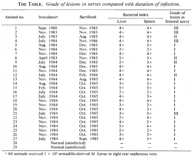
The total number of nine-banded armadillos studied was 29. They were provided by Dr. A. Dhople, Florida Institute of Technology, Melbourne, Florida, U.S.A. All had been inoculated with 108 M. leprae and sacrificed after disseminated disease became apparent. The duration of disease was 1½years in 22, 2 years in 4, and 5 years in 1. The remaining two normal armadillos served as controls (The Table). The femoral nerves from two levels of each animal were obtained at autopsy and were fixed in 10% neutral formalin. Each nerve specimen was divided into two parts, one for routine paraffin embedding (hematoxylin-eosin and Fite-Faraco staining), the other part being cut into many 1-mm3 pieces and refixed in 2.5% glutaraldehyde after rinsing in phosphate-buffered saline. Subsequently, the specimens were postfixed in 10% osmium tetraoxide, dehydrated, and embedded in Epon 812. Ultrathin sections were cut and stained with uranyl acetate and lead citrate. Finally, the stained grids were viewed with an H-600 electron microscope.
RESULTS
Light-microscopy observations. All animals showed lesions except the controls. The most remarkable sites of the lesions were in the loose connective tissue and fatty tissue between nerve bundles. All were marked by congestion and edema, infiltration with large numbers of macrophages and a few lymphocytes, occasional polymorphonuclear leukocytes (PMN) and even plasma cells. These macrophages harbored leprosy bacilli with globi but without prominent foamy cytoplasm. Although leprosy bacilli were not seen in the PMN, the lymphocytes, or the plasma cells, they were sometimes seen in fibroblasts, in the endothelial cells of small vessels (including capillaries or lymphatics), and in the smooth-muscle cells of the walls of venules. Sometimes there were macrophages containing leprosy bacilli in the lumen of small vessels. We observed in early lesions that there may be a perivascular accumulation of bacillated macrophages which gradually fuse together to form large sheets. These bacillated macrophages also infiltrated between fatty cells and among nearby muscle fibers. The lesions in the nerve bundles were usually far less prominent than those in the surrounding soft tissues. The lesions were divided into three grades according to their severity. Nineteen animals were classified as grade I, the lowest grade lesions. These lesions contained only one or a few foci of bacillated macrophages in the perinerium or even a few bacilli in the nearby nerve fibers. There were no remarkable changes inside the nerve fibers in these animals (Fig. 1). Four animals had grade II lesions, in which many small foci of proliferating Schwann cells were associated with a slight infiltration of lymphocytes. Fite-Faraco (FF) staining disclosed moderate amounts of leprosy bacilli in the nerve fibers. Four animals had grade III lesions, in which nerve fibers were severely and diffusely infected, with prominent proliferation of Schwann cells, and there was an infiltration of lymphocytes in numerous foci associated with macrophages. There were large numbers of leprosy bacilli forming globi or in a beaded arrangement in the vicinity of the Schwann cell nuclei, sometimes parallel to the longitudinal axis of the nerve fibers. No granular bacilli could be found. In the perineurium, there were inflammatory infiltrations with many bacilli but without fibrosis (Fig. 2).
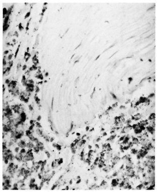
Fig. 1. Grade I lesion: Prominent perineurial infiltration of bacillated macrophages affecting focal arcas of perincurium in which bacilli could be found. No lesion is seen in nerve bundle (FF x 450).
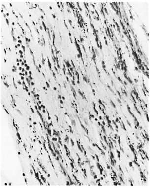
Fig. 2. Grade III lesion: Nerve bundle with diffuse lesions; bacilli are seen near the nuclei of many Schwann cells associated with perivascular lymphocytic infiltration (FF x 400).
The severity of the lesions in peripheral nerves in this series was closely related to the duration of the infection (The Table); 1 animal for 5 years, 2 for 2 years, and 1 for 1 year in grade III. The duration of infection in grade I and grade II was 1 year or a little more than 1 year, except for one animal for 2 years. These findings suggest that lesions in peripheral nerves of armadillos become prominent only after 2 years of infection.
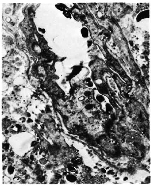
Electron-microscopic observations. There were large sheets of macrophages surrounding the peripheral nerves. Their arrangement was tightly packed and they contained abundant organelles in their cytoplasm, prominent Golgi apparatus, centrioles, cytoplasmic microfilaments, and mitochondria (Fig. 3). Occasionally clusters of glycogen granules inside mitochondria could be seen (Fig. 4). The rough endoplasmic reticulum (RER) was rather small, but there were many lysosomes containing leprosy bacilli forming phagolysosomes. The leprosy bacilli were intact and solid; aggregation of the cytoplasm of the bacilli was rarely visible. Most myelinated nerve fibers appeared to be normal, while unmyelinated nerve fibers were seriously affected (Fig. 5). Many unmyelinated axons contained leprosy bacilli in their superficial part embraced by thin, interrupted cytoplasmic membranes of the nearby Schwann cells (Figs. 6 and 7). The affected axoplasm became more rarefied, with neurofilaments being condensed or becoming indistinct (Fig. 8). Mitochondria in the axoplasm showed swelling with various sizes of vacuoles, the appearance of myelin figures, and even some myelin detritus. Most myelin sheath layers in myelinated nerve fibers remained intact except for some foci of loosening of the myelin layers and their protruding into the axon (Fig. 9). Fibroblasts of the endoncurium were marked by an abundance of RER and harbored bacilli. Sometimes leprosy bacilli would appear between collagen fibers, presumably arising from ruptured infected cells (Fig. 10).
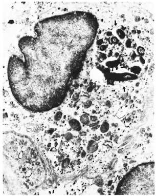
Fig. 3. Macrophage showing abundant cytoplasmic organelles and numerous lysosomes; (↑) = aggregate of bacilli inside phagosome with indistinct membrane ( x 12,000).
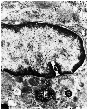
Fig. 4. Granules of glycogen inside mitochondria (G) and a bacillus (↑) in perinuclear space ( x 24,000).
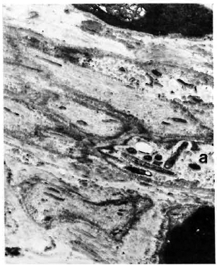
Fig. 5. Several axons (a) of unmyelinated nerve fibers; two axons contain bacilli with rarefaction of axoplasm ( x 6000).
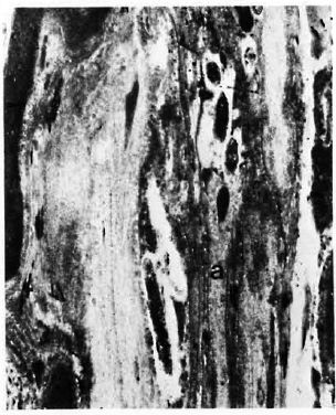
Fig. 6. Bacilli in axon (a) of unmyclinated nerve fibers with surrounding electron-transparent area ( x 8000).
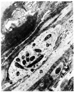
Fig. 7. Numerous bacilli in Schwann cells (S) of unmyelinated nerve fiber with indistinet membrane separating it from axon (a) below ( x 18,000).
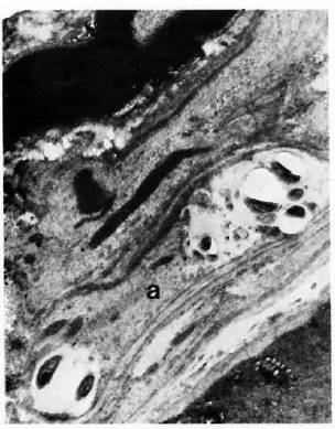
Fig. 8. Bacilli in axons (a) of unmyelinated nerve fibers; neurofilaments condensed between two clusters of bacilli ( x 10,000).
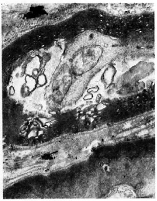
Fig. 9. Various sues of vesicles in axoplasm of myelinated nerve fibers and focal loosening of myclinated layers ( x 12,000).
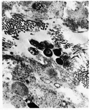
Fig. 10. Extra cellular bacilli between collagen fibers ( x 20,000).
Furthermore, there were infiltrations of the nerve with lymphocytes, plasma cells, and bacillated macrophages. Endothelial cells of the small vessels in nerve bundles showed swelling of the cytoplasm, pinocytosis, enlarged mitochondria, and some bacilli within the vessel lumen (Fig. 11). In bacillated cells there were many lysosomes near bacilli with electron-transparent areas surrounding the leprosy bacilli. Rarely, some dense bodies resembling the leprosy bacillus in cross-section appeared in the perinuclear space of some of the macrophages (Fig. 4). The endothelial cells of venules and capillaries were swollen, with vacuolization and enlarged mitochondria. Occasionally leprosy bacilli could be identified in endothelial cells. Similar changes to those seen in bacillated macrophages were also observed in perithelial cells of small vessels. Schwann cells showed cytoplasmic swelling, increased number of organelles, and bacilli in their cytoplasm. There were electron-transparent areas and granular substance surrounding bacilli which may participate in the formation of globi and become membrane bound.
11. Swollen endothelial cells of lymphatics with bacilli incide lumen of lymphatic vessel ( x 12,000).
DISCUSSION
The only mycobacterium which invades peripheral nerves is the leprosy bacillus, therefore the study of the lesions in peripheral nerves is important for understanding the pathogenesis of leprosy. Job (3) pointed out that Schwann cells are the target cells of the leprosy bacillus. In human lepromatous leprosy, the leprosy bacillus may be found in the Schwann cells, macrophages, endothelial and perithelial cells of small vessels, or even in the axon of the nerve fibers. There are only three reports on changes in the peripheral nerves of leprosy infected armadillos. Yoshizimi, et al. (8) and Balentine, et al. (1) concluded that lesions in the peripheral nerves of armadillos were much the same as human lepromatous lesions, except that no bacilli could be found inside the axons of the nerve fibers. Mehta and Antia (6) state that they found bacilli intra-axonally but, in our opinion, their photographs do not clearly demonstrate this finding.
We present in this paper the lesions of axons in detail, showing the formation of various sized vesicles, onion-skin myelin figures, and focal rarefaction or condensation of neurofilaments. A more important finding is the presence of leprosy bacilli in the axons of unmyelinated nerve fibers embraced by bacillated Schwann cells with interrupted cytoplasmic membranes which may rupture and liberate the bacilli into the axons. The lesions in axons are the results of the presence of bacilli. The fact that no bacilli could be found in the axons of myelinated nerve fibers may be due to the thick myelin sheath which separates the bacillated Schwann cells from the axons. One other important finding is the lack of regressive changes in lesions, such as fibrous proliferation or scar formation (5). This is in contrast to findings in neural lesions in human lepromatous patients and also to the findings of Mehta and Antia, who found increased endoneurial fibrosis in all of the nerves in the armadillo they observed. These findings suggest that the bacillated cells were disrupted much earlier or more easily in armadillos, with a high turnover and greater liberation of bacilli phagocytosed by the neighboring cells. The high turnover of these cells promotes the spread of lesions in nerves and explains the lack of a distinct foamy appearance of these cells.
In all the armadillos studied by us, the soft tissues adjacent to the perineurium were so prominently affected that these might be the primary lesions which then, subsequently, extended to the peripheral nerves. At first the perineurium was involved, later the lesions spread to the inner part of the nerve tissue, forming numerous focal lesions and, finally, the nerve became diffusely affected. The severity of the lesions is closely correlated with the duration of infection in the animals; the longer the duration of infection, the more severe the lesions. We divided the nerve lesions into three stages corresponding to different degrees of nerve damage. Fukunishi (2) induced lesions of peripheral nerves in nude mice by the injection of M. leprae. She concluded that the perineural lesions are the result of direct spread from the lesions of surrounding tissues.
The spread of lesions to the perineurium from soft tissues may be directly by migration of bacillated macrophages or the bacilli may pass through the lymphatics surrounding the nerves. These possibilities have been considered in human cases by Skinsnes (7). In our specimens, we found some lymphatics in the soft tissue lesions under lightand electron-microscopy. Their endothelial cells harbored bacilli, and some bacilli were found in their lumens. The findings substantiate the opinion that lesions of the peripheral nerves could spread by lymphatics. However, substantiation of lymphatic spread docs not preclude the possibility of spread via the hematogenous route. Endothelial cells and the pericytes of small blood vessels in nerve bundles can also harbor leprosy bacilli, indicating the presence of bacillemia. We, therefore, believe that lesions in peripheral nerves in leprosy may be initiated and spread by various routes.
Acknowledgment. We are deeply indebted to Dr. A. M. Dhople, Head of the Infectious Division, Florida Institute of Technology, Melbourne, Florida, U.S.A., for his kindness in supplying the spécimens.
REFERENCES
1. Balentine, J. D., Chang, S. C. and Issar, S. L. Infection of armadillos with Mycobacterium leprae; ultrastructural study of peripheral nerve. Arch. Pathol. Lab. Med. 100 (1976) 175-181.
2. Fukunishi, Y. Electron microscopic findings in the peripheral nerve lesions of nude mice inoculated with M. leprae. Int. J. Lepr. 53 (1985) 75-78.
3. JOB, C. K. Pathology of peripheral nerve lesions in lepromatous leprosy-a light and electron microscopic study. Int. J. Lepr. 39 (1971) 251-268.
4. Kirchheimer, W. F. and Storrs, E. E. Attempts to establish the armadillo (Dasypus novemcinctus, Linn.) as a model for the study of leprosy. I. Report of lepromatous leprosy in an experimentally infected armadillo. Int. J. Lepr. 39 (1971) 693-702.
5. Liu, T.-C, Qiu, J.-S. and Ho, Y.-F. Study on lesions of peripheral lepromatous nerves in 70 autopsied cases. Chin. J. Dermatol. 15 (1982) 149-152.
6. Mehta, L. and Antia, N. H. Ultrastructure of sciatic nerve of armadillo infected with Mycobacterium leprae. Indian J. Lepr. 56 (1984) 540-554.
7. Skinsnes, O. K. M. leprae and its "affinity"; for nerves. Int. J. Lepr. 39 (1971) 762-765.
8. Yoshizimi, M. O., Kirchheimer, W. F. and Asbury, A. K. A light and electron microscopic study of peripheral nerve in an armadillo with disseminated leprosy. Int. J. Lepr. 42 (1974) 251-259.
1. M.D., Professor and Chair, Research Department of Pathology;
2. M.D., Professor of Pathology, Director of Department of Electron Microscopy;
3. M.D., Ph.D., Honorary Professor of Pathology, Department of Pathology, Sun Yat-Sen University of Medical Science, Zhongshan Road, Guangzhou, People's Republic of China.
Received for publication on 13 June 1988.
Accepted for publication in revised form on 21 October 1988.