- Volume 67 , Number 2
- Page: 154–8
Effect of oil of hydnocarpus on wound healing
ABSTRACT
Oil of hydnocarpus has been replaced by other chemotherapeutic agents which have a better mycobactericidal effect. However, none of the currently used antileprosy drugs has been reported to have a positive effect in wound healing. Anecdotal reports claim that leprosy patients who have taken capsules containing oil of hydnocarpus orally have shown more rapid wound healing than those not receiving it. In view of these reports, a pilot experimental study was undertaken to determine the effect of the oil of hydnocarpus in wounds experimentally inflicted on male Wistar rats. The wound-healing effect of oil of hydnocarpus was studied with reference to collagenation and the strength of the scar tissue. The drug-treated group showed a significant increase in body weight and strength of scar tissue in the incision model and, also, increased strength of the collagen tissue and hydroxy-proline content in the dead space model. The results of this pilot study indicate that the oil of hydnocarpus, which also has antileprotic activity, could be a useful adjunct in the healing of wounds and ulcers in leprosy patients.RÉSUMÉ
L'huile d'hydnocarpus a été remplaçée par d'autre agents chimiothérapeutiques qui ont un meilleur effet mycobactéricide. Cependant, il n'y a pas de médicament contre la lèpre de décrit ayant une activité bénéfique sur la cicatrisation. Quelques rares rapports font état de cas isolé de cicatrisation plus rapide chez des patients atteints de lèpre ayant été traités par des capsules contenant de l'huile d'hydnocarpus comparé à des patients n'en ayant pas pris. Du fait de ces rapports, une étude pilote expérimentale a été entreprise pour déterminer les effets de l'huile d'hydnocarpus sur la cicatrisation de plaies induites expérimentalement chez des rats de souche Wistar. Le possible effet cicatrisant de l'huile d'hydnocarpus fut étudié en mesurant le degré de collagénisation et la résistance du tissu cicatriciel. Le groupe traité a montré une augmentation du poids corporel et de la résistance du tissu cicatriciel pour le modèle de plaie par incision, ainsi qu'une augmentation de la résistance du tissu collagénique et de la teneur en hydroxyproline pour le modèle de cicatrisation par seconde intention. Les résultats de cette étude pilote indiquent que l'huile d'hydnocarpus, qui présente également une activité antilépreuse, pourrait être un traitement adjuvant utile pour la cicatrisation des blessures et ulcères des patients lépreux.RESUMEN
En la actualidad, el aceite de hydnocarpus ha sido reemplazado por otros agentes quimioterapéuticos con mayor efecto bactericida. Sin embargo, no se ha reportado que alguna de las drogas antileprosas de uso actual tenga algún efecto curativo de las heridas de los pacientes con lepra. Algunos reportes anecdóticos ase-guran que los pacientes con lepra que han tomado cápsulas con aceite de hydnocarpus muestran una curación más rápida de sus heridas que los pacientes que no han tomado las cápsulas. En vista de estos reportes se hizo un estúdio piloto para determinar el efecto del aceite de hydnocarpus en heridas provocadas en ratas Wistar macho. El efecto curativo dei aceite de hydnocarpus se estableció en función de Ia colagenación y la firmeza del tejido cicatricial. El grupo tratado con la droga mostro un aumento significativo en el peso corporal y en la firmeza del tejido cicatricial en el modelo de incisión y un aumento en la firmeza del tejido colágeno y en el contenido de hidroxiprolina en el modelo del espacio muerto. Los resultados de este estúdio piloto indican que el aceite de hydnocarpus, el cual también tiene actividad antileprosa, podría ser útil en la cicatrización de las heridas y ulcéras en los pacientes con lepra.The phases of wound healing-collagenation, wound contraction and epithelization-are intimately interlinked. Collagen synthesis is a major aspect of wound healing. It is the process of collagen formation that gives the ultimate strength to the healed wound. Since collagen is rich in the amino acids hydroxyproline and hydroxylysine, of which hydroxyproline is predominant, estimation of the hydroxyproline content is a direct indicator of the collagen content in the granulation tissue. During collagen synthesis proline is incorporated into the polypeptide chains of pro-collagen within the fibroblast cells and hydroxylated by proline hydroxylase and molecular oxygen.
The oil of Hydnocarpus wightiana has been recognized for several years, as an antileprosy drug, as an acaracide and as an antiparasitic drug in the treatment of guinea worm infestations (10). The commercial sources of the oil are from Taraktogenos kurzii, Hydnocarpus wightiana and Hydnocarpus anthelminthica . However, more recent drugs have been developed which have a better safety profile and which are generally considered to have a better efficacy against most of these diseases.
In the treatment of leprosy none of the currently used antileprosy drugs has been reported to have a positive effect in the healing of wounds that are common in leprosy patients. However, recent anecdotal reports (10th Annual Report of the Samidha Charitable Trust, 1996: Manjrekar, S., p. 10 and Bagga, V. S., p. 4) claim that wounds in leprosy patients and in patients with diabetic ulcers and gangrene healed faster when the oil of hydnocarpus was given orally or administered topically. In view of these anecdotal reports, a pilot study was done to assess the effect of the oil of hydnocarpus on wound healing. The aim of the study was to evaluate the effect of the oil of hydnocarpus on the collagenation phase of wound healing.
MATERIALS AND METHODS
Twenty individually housed male rates of the Wistar strain were divided into two groups of 10 each. The animals in Group A served as controls. The animals in Group B were administered the oil of hydnocarpus at a dosage of 45 mg/kg/day, emulsified in gum acacia. None of the animals was put on any other medication.
Incision wounds. All of the animals were anesthetized using 35 mg/kg pentobarbital (pentobarbitone) administered in-traperitoneally. Following the method of Ehrlich and Hunt (4) a paravertebral incision 6 cm in length was made through the entire thickness of the skin at least 1 cm lateral to the vertebral column of each animal and the incision was closed with 4-0 silk sutures spaced 1 cm apart from each other.
On the tenth day, the sutures were removed and the breaking strength of the wounds was measured following the procedure of Lee (5). The animals were again anesthetized with 35 mg/kg pentabarbital and were secured to the operating table. A line was drawn on the skin at a distance of 3 mm on either side of the incision wound. Two Allis forceps facing each other were firmly applied on the line. On one side the forceps was hooked firmly to a metal rod fixed to the operating table. The other forceps was connected to a leak-proof, graduated polypropylene bottle through a string running over a pulley. The bottle was connected to a large water reservoir, placed at a suitable height, through a rubber tube which was kept occluded. To measure the breaking strength of the wound the tube was released to allow a constant flow of water from the reservoir into the graduated bottle, thus gradually increasing its weight so as to act as a pulling force to disrupt the wound. As soon as any wound dehiscence was observed, the rubber tube was clamped and the amount of water collected in the graduated tube was measured. Four readings were recorded for each incision. The mean of the four readings was taken as the breaking strength for the tissue.
Dead space wounds. These wounds were created by implanting polypropylene tubes of 2.5 cm x 0.5 cm subcutaneously in the dorsal aspect of the animals to harvest the granulation tissue, following the procedure described by Patil and Kulkarni (9). On the tenth day these polypropylene tubes were removed and the granulation tissue harvested around the tubes was dissected. The tubular granulation was cut longitudinally into two halves and each piece was analyzed separately for breaking strength as described above (5).
Determination of hydroxyproline content. The granulation tissue harvested from the dead space model was dried at 60°C for 24 hr in order to get the dry weight. The determination of hydroxyproline content followed the procedure of Neuman and Logan (7). The tissue was place in a sealed tube containing 10 ml of 6 N hydrochloric acid and hydrolyzed by heating at 110°C for 24 hr. The hydrosylate was cooled and any excess acid was neutralized with 10 N sodium hydroxide, using methyl red as an indicator. The volume of neutral hydrosylate was made up to 20 ml with distilled water.
From this, 0.1 ml of the hydrosylate was pipetted into clean, dry tubes and the volume made up to 0.5 ml using distilled water. From the stock solution of standard hydroxyproline, 1.6 ml was taken and diluted up to 100 ml, of which 0.5 ml (8 µg) was taken. To the above solution were added 1 ml each of 2.5 N NaOH, 0.01 M CuS04 and 6% H2,02,. The test tubes were immediately placed in a water bath at 80°C for 16 min and then cooled for 5 min. To this solution were added 2 ml of a freshly prepared solution of 5% para-dimethyl amino benzaldehyde in N-propanol and 4 ml of 3 N H2,02,. The tubes were again placed in the water bath at 80° for 15 min and then cooled for 5 min. For the estimation of hydroxyproline, the optical densities of the pink color of these test samples were compared to that of standard hydroxyproline of a known concentration at 540 nm using a Beckman Du-64 spectrophotometer.
RESULTS
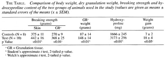
Incision wound model. In all of the animals the wounds healed well without any secondary infection. The linear scar tissue which had formed was indicative of good wound healing by primary intention. The breaking strength of the scar tissue in the test group was significantly greater than that of the control group (The Table), showing that the healing of the wound in the test group was significantly better.
Dead space model. The polypropylene tubes which acted as inert foreign bodies within the animal had induced good formation of granulation tissue in all of the animals. The breaking strength of the granulation tissue formed in the test group was higher than that in the control group, indicating better collagenation of the tissue in the test group. This was also confirmed by measuring the hydroxyproline content of the granulation tissue harvested from the dead space.
Hydroxyproline content. Both groups showed good collagenation as evidenced by the hydroxyproline content in the granulation tissue, but the hydroxyproline content of the granulation tissue in the test group was significantly higher than in the control group (The Table).
Two animals in the control group succumbed to the procedure on day one, and were excluded from the study. All of the other animals of both groups fed well and showed no alteration in feeding or fluid intake. The animals were quite mobile in their cages with no limitation of activity or signs of morbidity. All of the animals showed an increase in body weight but there was a significant increase in weight gain in the group that received the oil of hydnocarpus (The Table).
DISCUSSION
The oil extracted from Hydnocarpus wightiana was used in this study because it is the only species of the Hydnocarpus that is known to grow in this geographical location. The amber-colored oil of hydnocarpus had been used for the treatment of leprosy, being administered topically, orally and parenterally. It probably had little effect on established lepromatous leprosy but, by encouraging resolution, it may have prevented some indeterminate and early borderline forms from progressing into lepromatous leprosy (3). Although largely replaced by newer drugs, hydnocarpus oil is still employed in the treatment of leprosy in areas where it is cheap and readily available (6). The oils obtained from the Hydnocarpus -related species are therapeutically indistinguishable. Preparation of a pharmaceutical grade of the oil requires pressure extraction of the crude oils from the seeds, followed by saponification with sodium hydroxide. Repeated washings with hot water allow a purer oil to be decanted each time (8).
The oil of hydnocarpus contains fatty acids such as chaulmoogric acid, which has 18 carbon atoms, and hydnocarpic acid, which has 16 carbon atoms and their glycerides (10). These acids differ slightly in their chemical composition and optical rotary powers (8). Ethyl esters, cinnamates, beta-glycerophosphates and sodium salts of hydnocarpic and chaulmoogric acids have been found to be effective when applied topically (10).
It was suggested that the introduction of the oil of hydnocarpus, a foreign lipid, activates host lipases to destroy all foreign lipids including both the oil itself and the cell wall of the mycobacterium. One hypothesis held that once the bacillary wall was penetrated regular immunological processes could destroy the bacillus. Another hypothesis invoked counter irritation which resulted in chemotaxis; the irritation being caused by injection of the oil drew phagocytes into the vicinity of the bacillus (1-2).
An analysis of the chemical structure of these oils shows that they contain fatty acids which have a cyclopentene ring at the end of a carbon chain and an asymmetric carbon atom. Chaulmoogric acid is isomeric with linoleic acid but, unlike other fatty acids which have an open chain, chaulmoogric acid has a closed chain. Gor-lic acid is another long-chain fatty acid found in the oil of hydnocarpus, but its exact role is not known. The acids also contain a double bond in the cyclopentenyl ring. There is, consequently, a high possibility that both hydnocarpic acid and chaulmoogric acid could be free radical scavengers, a property which could contribute to the pro-healing effect seen in the study. Being long-chain carboxylic acids, they are lipophilic and cause dissolution of the my-colic acid coat of the mycobacteria, thereby causing cell death.
CONCLUSION
It is commonly believed that the oil of hydnocarpus and its derivatives are of little or no curative value, and their unpleasant side effects probably outweigh any advantage that may accrue from their use (s). As a result, it has been dropped from most formularies and memories. However, no well-designed clinical (or experimental) trial of the oil was ever conducted. From the present study, it could be suggested that administration of the oil orally would trigger the synthesis and secretion of collagen from the fibroblast cells to give a better strength to the healing wound and the scar tissue. At the given dosage, the drug-treated group showed a significant increase in body weight, strength of scar tissue in the incision model and, also, increased strength of the collagen tissue and hydroxyproline content in the dead space model.
If the dosage that was used for oral administration in this study is extrapolated to clinical trials, the untoward gastrointestinal side effects that were seen in the past might be avoided while the beneficial effects of the oil in promoting wound healing could be utilized. The results of this pilot study indicate that the oil of hydnocarpus when administered orally, with its already known antileprotic activity, could be a useful adjunct in the treatment of leprosy. Further studies with regard to its wound-healing properties are in progress.
Acknowledgment. The authors wish to express their gratitude to M/s. Samidha Charitable Trust, Mumbai, India, for the generous supply of the oil of hydnocarpus used in the study; to Prof. K. S. Karanth. Head, Department of Pharmacology, Kasturba Medical College, Manipal. India, for the use of the department facilities, and to Dr. Gopalan Kutty of the same department for critical evaluation of the pharmaceutics of the oil.
REFERENCES
1. Cochrane, R. G. A Practical Textbook of Leprosy. Oxford: Oxford University Press, 1947, pp. 117-132 (cited in Norton).
2. Cole, H. I. Chemistry of antileprosy drugs. Int. J. Lepr. 1(1932)159-164 (cited in Norton).
3. Davey, T. F. Common features in rapidly declining leprosy epidemics. (Editorial) Lepr. Rev. 46(1975)5-8.
4. Ehrlich, H. P. and Hunt, T. K. Effect of cortisone and anabolic steroids on tensile strength of healing wounds. Ann. Surg. 170(1969) 203-206.
5. Lee, K. H. Studies on the mechanism of action of salicylates: retardation of wound healing by aspirin. J. Pharmacol. Sci. 57(1968) 1042-1043.
6. Martindale, W. The Extra Pharmacopoeia . 27th ed. Reynolds, J. E. F., et at ., eds. London: Pharmaceutical Press, 1979, p. 1504.
7. Neuman, R. E. and Logan, M. A. The determination of collagen and elastin in tissue. J. Biochem. 186(1950)549-552.
8. Norton, S. A. Useful plants of dermatology. I. Hydnocarpus and chaulmoogra. J. Am. Acad. Dermatol. 31(1994) 683-686.
9. Path., P. A. and Kulkarni, D. R. The effect of antiproliferative agents on healing of dead space wounds in rats. Indian J. Med. Res. 79(1984) 445-447.
10. Pharmaceutical Society of Great Britain. The British Pharmaceutic Codex . London: Pharmaceutical Press, 1934, pp. 710-711.
1. B.Sc., M.D.; Department of Pharmacology. Kasturba Medical College, Manipal 576119, India.
2. M. Pharm., Ph.D.; Department of Pharmacology. Kasturba Medical College, Manipal 576119, India.
3. M. Pharm., Department of Pharmacology. Kasturba Medical College, Manipal 576119, India.
Reprint requests to Dr. Oommen at the above address or FAX 91 -8252-70062.
Received for publication on 24 September 1998.
Accepted for publication in revised form on 5 February 1999.