- Volume 65 , Number 1
- Page: 97–99
Influence of DDT exposure on susceptibility to human leprosy bacilli in mice
To the editor:
The widespread use of 1,1,1 -trichloro-2,2-bis (p-chlorophenyl) ethane (DDT) over a number of years, coupled with its extreme stability and slow metabolism, has led to environmental contamination and the ultimate carryover from the food into humans (2,4). Subchronic exposure to DDT perturbs immune responsiveness and, hence, may alter susceptibility of the host to pathogens. The increase in susceptibility to infection with Histomonas meleagridis and the hepatitis virus in birds exposed to chronic doses of DDT has been reported. All of the above findings have been reviewed by us recently (1). Since leprosy is a priority socioeconomic and public health problem in many of the developing countries, it is appropriate to study the influence of DDT on the susceptibility of the host to experimental infection of Mycobacterium leprae. With this in mind, the present study was designed to evaluate the subchronic effect of DDT on the growth of M. leprae in the mouse foot pad.
Pure p,p'- DDT was obtained by repeated crystallization of the technical grade material (Hindustan Insecticide Ltd., India) from 95% ethanol until it showed a single peak by gas chromatography. Albino male mice (Rockfeller strain) weighing 18-20 g were maintained under standard laboratory conditions and provided with a diet containing 0 (control), 20, 50 or 100 ppm of p,p'-DDT and water ad libitum throughout the experiment. Each treatment group consisted of 18-22 mice.
Mice (8-12 group) were inoculated in the right hind foot pad with 7.5 x 10³ bacilli in 0.03 ml Hanks' balanced salt solution (HBSS) containing 0.1% bovine serum albumin (BSA) obtained from human biopsy samples. Ten mice from each treatment group were inoculated with 0.03 ml of medium. The exposure to DDT was commenced from the day of inoculation. The mice were killed and the bacilli were harvested from the foot pads after 24 weeks of DDT exposure.
Harvesting was done according to the method of Desikan and Venkataramanaiah (3) with suitable modifications. The foot was cut off above the ankle joint, washed thoroughly with soapy water, and rinsed. The foot pads were separated and homogenized individually in chloroform. The supernatant was collected and the chloroform was removed by evaporation at 37ºC. The residue was suspended in 1 ml HBSS with 0.1% BSA. The smears were prepared in triplicate from a fixed volume of suspension and were stained by the Ziehl-Neelsen method for acid-fast bacilli (AFB). The total number of bacilli per foot pad was calculated. The data were analyzed using the Mann-Whitney U test (two-tailed). A p value of at least 0.05 was considered to be the level of significance in all statistical tests.
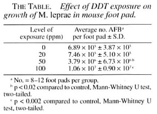
The growth of M. leprae as assessed from the AFB count in the mouse foot pad after 24 weeks of inoculation is shown in The Table. The data reveal that the growth of M leprae is significantly (p < 0.02) enhanced in mice exposed to 50 or 100 ppm DDT in a dose-dependent manner in comparison to growth in the controls. DDT exposure at the 20-ppm level did not significantly alter the growth rate of leprosy bacilli in the mouse foot pad. No bacilli were observed in mice inoculated with 0.03 ml of medium.
There is no information available on the possibility of growth of leprosy bacilli in DDT-exposed subjects. Admittedly, high levels of DDT and its metabolites have been reported in the body fat (0.32 to 380 ppm) and blood (0.02 to 4.61 ppm) of humans (4). DDT administration in experimental animals through food accumulates in the body fat and blood, and the body load can be compared with human concentrations which could result from environmental contaminations (2). Hence, it has been considered desirable to investigate the susceptibility to M. leprae infection in DDTexposed mice.
Enhanced bacillary growth in DDT-exposed mice demonstrates that DDT increased the susceptibility to leprosy infection in a dose-dependent manner. It is well established that resistance to infection by M. leprae is conferred by cell-mediated immunity, and in the immunocompromised (thymectomized and gamma-irradiated) animal model, an enhanced growth of M. leprae has been reported (5). Our previous studies have shown that the subchronic administration of DDT produces marked suppression of cell-mediated and humoral immune responses in experimental animals in a dose-dependent pattern (1). Hence, altered susceptibility of DDT-exposed mice to M. leprae infection might be attributed to DDT-induced immune dysfunction. Although it appears that the immunosuppressive effects of DDT could be one of the factors for increased growth of M. leprae in normal mice, it is not known whether the pesticide itself could also enhance the multiplication of the bacilli. More light can be thrown in this direction by in vitro studies of the effect of DDT on the growth of M. leprae- related mycobacteria, peritoneal macrophage activity, and the release of interleukin - 2 by T cells.
Testing of the effects of DDT on host resistance is important in relation to the health aspects of pesticides, particularly due to the widespread use of DDT and its persistence in the environment. It is apparent that a more complete understanding of the toxicity of DDT is necessary in order to determine the human health hazards and to establish guidelines for acceptable DDT residues in the environment. Adverse effects of DDT on immune function could place the host in a more vulnerable position regarding various pathogens. There is a need for further detailed studies on the dose-time relationship of DDT exposure and the growth of M. leprae in mice since repeated exposure and contamination are possible in nature.
- Basu D. Banerjee, M.Phil., Ph.D.
Bidhan C. Koner, M.D.
Department of Biochemistry
University College of Medical Sciences& G.T.B. Hospital
(University of Delhi)
Shahdara
Delhi 110095, India
- Sayed T. Pasha, M.Phil., Ph.D.
Department of Biochemistry
National Institute of Communicable Diseases
Delhi 110054, India
Acknowledgment. The authors are indebted to the kite Professor S. Chaudhuri, School of Tropical Medicine, Calcutta, and Dr. K. K. Dutta. Director, NICD. Delhi, for their keen interest and valuable help during the course of study.
REFERENCES
1. BANERJEE, B. D., KONER, B. C. and RAY, A. Immunotoxicity of pesticides: perspectives and trends. Indian J. Exp. Biol. 34(19%)723-733.
2. BANERJEE, B. D., PASH. S. T, GULATI, M. and HUSSAIN, Q. Z. DDT and its metabolites in body fat and liver of albino mice-biochemical and histological studies. J. Com. Dis. 14(1982)274-280.
3. DESIKAN, K. V. and VENKATARAMANAIAH, H. N. A modified method of harvesting M. leprae from foot pads of mice. Lepr. India 48(1976)157-162.
4. RAMCHANDRAN, M., BANERJEE, B. D., GULATI, M., GROVER, A., ZAIDI, S. S. A. and HUSSAIN, Q. Z. DDT and HCH residues in the body fat and blood samples from some Delhi hospitals. Indian J. Med. Res. 80(1984)590-593.
5. TALWAR, G. P., TURK, J. L. and REES, R. J. W. Progress in immunology of leprosy. In: Growth of the ICRC Bacilli in the Foot-Pad of Mice. Bapat, C. V. and Modak, S., eds. New Delhi: Arnold-Heinemann Publishers Pvt. Ltd.. 1983. pp. 31-42.