- Volume 65 , Number 2
- Page: 166–9
Auditory brain stem evoked potentials in patients with leprosy
ABSTRACT
Nineteen, randomly selected male patients with lepromatous leprosy were evaluated electrophysiologically. All of these patients had long-standing disease and were treated with dapsone alone. There were statistically significant differences between the values obtained in this group of leprosy patients compared to 20 age-matched controls in auditory brain stem evoked potentials (ABEP). The findings are consistent with a pathologic process located mainly between the cochlear nucleus and the lateral lemniscus in the auditory brain stem pathways. It should be emphasized that our patients had long-standing disease which was treated with dapsone. ABEP could very well be different in leprosy patients diagnosed early and treated for relatively short periods with multidrug therapy. Brain stem evoked response audiometry may be useful for evaluating the possibility of brain stem involvement in leprosy.RÉSUMÉ
On a évalue du point de vue électro-physiologique 19 patients masculins atteints de lèpre lépromateuse et sélectionnés de manière aléatoire. Tous ces patients avaient une maladie de longue durée et étaient traités par dapsone en monothérapie. 11 y avait des différences statistiquement significatives entre les valeurs obtenues dans ce groupe tie malades de la lèpre, comparés à 20 témoins appariés pour l'âge, en ce qui concerne les potentiels cérébraux auditifs évoqués ( PC Ali). Ces observations sont cohérentes avec un processus pathologique localisé principalement entre le noyau cochléaire et le lemnisque latéral dans les voies de conduction auditive cérébrale. Il faut insister sur le fait que nos patients avaient une maladie de longue durée qui était traitée à la dapsone. Les PCAE pourraient très bien être différents chez des malades de la lèpre diagnostiqués précocement et traités par polychimiothérapie. L'audiométrie de la réponse évoquée cérébrale pourrait être utile pour évoquer la possibilité d'une implication cérébrale dans la lèpre.RESUMEN
Se hicieron estudios electrofisiológieos en 19 pacientes masculinos con lepra lepromatosa seleccionados al azar. Todos los pacientes tenínan una enfermedad de larga duración y fueron tratados sólo con dapsona. Hubieron diferencias estadísticamente significativas en los potenciales auditivos evocados del tronco cerebral (PAETC) entre los pacientes con lepra y los controles (20) de la misma edad. Los hallazgos son consistentes con un proceso patológico localizado principalmente entre el núcleo coclear y el lemniscus lateral de las vías auditivas del tronco cerebral. Debe enfatizarse que nuestros pacientes tuvieron una enfermedad de larga duración tratada sólo con dapsona. Los patenciaies auditivos evocados del tronco cerebral pudieron haber sido diferentes, si los pacientes con lepra hubieran sido diagnosticados tempranamente y tratados con poliquimioterapia durante periodos más cortos de tiempo. La medición de la respuesta auditiva evocada del tronco cerebral puede ser útil para evaluar la posibilidad de afección del tronco nervioso en la lepra.Leprosy is a chronic, systemic, infectious disease caused by Mycobacterium leprae. It manifests itself as specific granulomatous or neurotrophic lesions in the skin, mucous membranes, nerves, eyes, bones, and viscera. Central nervous system involvement in leprosy is still debated (11,12,20).
Conventional investigations on auditory system involvement in leprosy have revealed contradictory results. Decandia and Mariano reported specific involvement of the cochlea and acoustic nerve in leprosy (5). Mann, et al. reported cochlear-type hearing loss (13). However, Cochrane stated that the acoustic nerve is never affected in leprosy and there are no known lesions in the middle or inner ear (3). Sensorineural hearing loss in leprosy was observed by El Arini, et al., and they concluded that this hearing loss was due to the involvement of the acoustic nerve (7). Chehata reported the retrocochlear origin of the sensorineural hearing loss of leprosy patients (2). Singh, et al. have reported cochlear hearing loss, and they concluded that acoustic nerve involvement seems to be unlikely (20).
Auditory brain stem evoked potentials (ABEP) is a noninvasive and reliable electrophysiologic method for the assessment of the brain stem structures traversed by auditory pathways. Our electrophysiologic study was designed to investigate the involvement of the central auditory pathways in leprosy.
MATERIALS AND METHODS
Thirty-eight ears of 19 randomly selected male patients with lepromatous leprosy were evaluated for this study. They were compared with 20 age-matched male controls. Subjects with a past history of ear disease, exposure to excessive noise, systemic illness (diabetes mellitus, hypertension, renal failure), ototoxic drugs, head or ear trauma, or a family history of deafness were excluded From the study.
Otorhinolaryngological examinations were performed by the same otorhinolaryngologist (OC), and all audiological tests were performed by the same tester (AO). The informed consent of all subjects was obtained before clinical and audiometric examinations.
Pure tone audiometry was performed using an Interacoustics Clinical Computer Audiometer (Model AC5; Interacoustics Co., Assens, Denmark) in a soundproof room (Industrial Acoustics Co., Bronx, New York, U.S.A.). Both air and bone conduction were tested (250-8000 Hz). Compatible TDH-39P earphones mounted in Telephonies cushions and an Oticon 62507 bone vibrator (Oticon International A/S, Copenhagen, Denmark) were utilized.
ABEP were recorded in a soundproof semidark room using a Medelec Audiostar Electric Response Audiometer (Vickers Healthcare Co., Surrey, England). Silversilver chloride surface disk electrodes were placed on the vertex (active), the ipsilateral mastoid (negative) and the contralateral mastoid (ground) areas of the head. The electrode impedance was maintained below 2000 ohms. Auditory brain stem potentials were evoked by a 0.1 ms rarefaction click stimulus. The stimulus was presented monaurally through TDH-49P earphones (mounted in Telephonies cushions) at repetition rates of 10 and 50 sps and at 80 dB nHL. Two sets of 1024 responses, with a recording bandpass of From 200 to 3000 Hz (3 dB down points, 6 dB/octave), were averaged for each ear and recorded on an X-Y plotter. The replicability of recordings was determined by comparing two trials. The absolute latencies of waves I, III and V and the interpeak latencies of waves I-III, III-V and I-V were measured and calculated.
Statistical analyses were performed using the StatView 512+® program (Brainpower, Inc., Calabasas, California, U.S.A.). The data are expressed as mean ± S.D. Comparison of the two groups was performed with Student's t test.
RESULTS
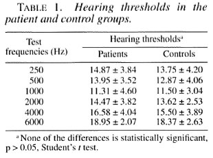
Nineteen males with leprosy (38 ears) aged between 33-59 years (average age 45.3 ± 8.9 years) were compared with 20 male controls (40 ears) aged between 35-60 years (average age 46.8 ± 8.2 years). Neither the leprosy patients nor the controls showed more than a 20 dB HL hearing threshold at any of the test frequencies (Table 1).
Duration of the disease was 10-42 years (mean 26.7 ± 9.3 years). The age-at-onset was 9-36 years (mean 20.6 ± 7.7 years).
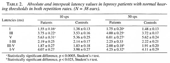
The mean latency values for all leprosy patients and control subjects were calcu lated and statistically analyzed. No differ ences between the right and left ears were observed (p > 0.05). All absolute latencies (waves I, III, and V) at both repetition rates (10 and 50 sps) (p < 0.0005) and the III-V interpeak latency at 50 sps (p < 0.025) of the leprosy patients were significantly longer than those of the control subjects (Table 2).
DISCUSSION
In an ABEP study on 47 leprosy patients, abnormal latency values were detected in only three cases (6%) and the mean ABEP interpeak latencies did not differ between the leprosy and control groups (1).
In our study, the absolute latency values derived From the patients using low and high repetition rates were increased com pared with the controls. Nevertheless, only the interpeak latency values between waves III and V and waves and I and V were sig nificantly (p < 0.025) increased for the high rate data obtained From patients. The values of the I-III interpeak latency were not dif ferent From those derived From the controls. In light of these results, it would be reason able to state that the abnormality of the in creased absolute latency values was mostly the result of a delay in wave I, and the increased III-V interpeak latency was mostly the result of a delay in wave V. Observing the increases in absolute latency of waves I and V, and the interpeak latency of wave III-V, besides having normal values for other interpeak latencies, implies that this result is consistent with a pathology mainly limited to the brain stem rather than to the cochlea. Influence on the central auditory pathways is evident between the cochlear nucleus and the lateral lemniscus, but this influence probably also exists in the auditory nerve. An increase in the III-V interpeak latency appeared only at a repetition rate of 50 sps. The effect of the increased stimulus rate on ABEP has been attributed to central synaptic changes (17,18) Eggermont and Schmidt stated that when the interpeak interval for I-III is normal but increased for III-V, the lesion is likely to be located in the upper or middle pons and might be extending into the caudal midbrain (6).
The pathologic process located in the pons probably would be related to oligodentrocytes rather than to the nerve tracts, and this relates to the findings of normal hearing thresholds but abnormal evoked potential latency values. Ortega, et al. noted that Mycobacterium leprae were contained within the sacuoles and appeared in the macrophages and Schwann cells in both lepromatous and borderline leprosy (16). These ultrastructural changes were observed in the peripheral nervous system. Schwann cells in the peripheral nervous system and oligodentrocytes in the central nervous system insulate axons by forming a myelin sheath, which greatly enhances the conduction of electrical signals (9). Thus, affecting of myelin sheath in leprosy is probably the underlying reason for the desynchrony of the neural discharges which lead to abnormality in the evoked potential recording without any detected hearing loss.
Patients received dapsone for their therapy. Since there is no data about an ototoxic adverse effect of this drug in the literature, we may conclude that leprosy rather than the drug accounts for the abnormality in the evoked potentials in patients with leprosy. All of the patients investigated in this study had long-standing disease and were treated with dapsone alone. In 1982, the World Health Organization recommended multidrug therapy (WHO/MDT) for the treatment of leprosy and, as a result, monotherapy with dapsone has given way to MDT since that time (21,22). Leprosy patients are treated with MDT more effectively (8,10,14,15,19). MDT has shortened the course of treatment and reduced deformity rates (4,14). Investigation of ABEP variations in such treated cases should be the subject of a new study.
Acknowledgment. The authors would like to thank Biilent Serbetcioglu, M.D., Ph.D. for reviewing the English of the manuscript.
REFERENCES
1. BARTEL., P. R.. BAKER, M. K.. COMBRINK, P., ROBINSON, E. and VAN DER MEYDEN, C. H. Auditory brainstem evoked potentials in leprosy. S. Afr. Med. J. 73 (1988) 593-595.
2. CHEHATA, M. L'atteinte du nerf auditif dans la lèpre. Ann. Oto. Laryng. (Paris) 95 (1978) 685-690.
3. COCHRANE-, R. G. Lesions of the nose, ear, mouth and throat. In: Leprosy in Theory and Practice. 2nd edn. Cochrane, R. G. and Davey, T. F., eds. Baltimore: Williams and Wilkins Co., 1964, pp. 322-326.
4. COURTRIGHT, P., LEWALLEN, S., LI, H. Y., HU, L. F. and YANG, J. W. Lagophthalmos in a multibacillary population under multidrug therapy in the People's Republic of China. Lepr. Rev. 66 (1995) 214-219.
5. DECANDIA, A. and MARIANO, A. Studies of cochleovestibular function in patients with leprosy. Riv. Audiol. Prat. 1 (1960) 135-138. (quoted in 5).
6. EGGERMONT, J. J. and SCHMIDT, P. H. The auditory brainstem response. In: Evoked Potential Manual. Colon, E. J. and Visser, S. L., eds. Antwerp: Kluwer Academic Publications, 1990, pp. 41-77.
7. EI. ARINI, F., SHITIATA, M. A. and Anon ZEID, S. A. Eighth cranial nerve affection in leprosy. Int. J. Lepr. 38(1970) 164-169.
8. FREERKSEN, E. Preliminary experience with combined therapy using rifampicin and isoprodian. Lepr. Rev. 46 Suppl. (1975) 161-163.
9. KANDEL, E. R. Nerve cells and behavior. In: Principles of Neural Science. 3rd edn. Kandel, E. R.. Schwartz, J. II. and Jessell, T. M.. eds. New York: Elsevier, 1991, pp. 18-32.
10. KATOCH, K., NATRAJAN. M., YADAV. V. S. and BHATIA, A. S. Response of leprosy patients with single lesions to MDT. Acta Leprol. 9 (1995) 133-137.
11. KATOCH, K., RAMU, G., SENGUPTA, U. and BHARADWAJ, V. P. Central nervous system involvement in leprosy. Indian J. Lepr. 56 (1984) 813-818.
12. KOYUNCU, M., CELIK, O.. ÖOZTÜRK A. and SAUNDERS, M. Audiovestibular system, fifth and seventh cranial nerve involvement in leprosy. Indian J. Lepr. 66(1994) 421-428.
13. MANN, S. B. S., KUMAR, B, YANDE. R., KAUR. S, KAUR, I. and MEHRA, Y. N. Eighth nerve evaluation in leprosy. Indian J. Lepr. 59 (1987) 20-25.
14. MCDOUGALL, A. C. Implementing multidrug therapy for leprosy. Oxford: Oxfam, 1988. Oxfam practical guide no. 3.
15. NOORDEEN, S. K. Elimination of leprosy as a public health problem: progress and prospects. Bull. WHO 73 (1995) 1-6.
16. ORTEGA, V. V, MARTINEZ-DIAZ, F., ORTUNO-PACHECO, G. and CALDERAN-RUBIALES, F. A. P. Ultrastructural study across the leprosy spectrum. Ultrastruct. Pathol. 18(1994)423-432.
17. PRATT, H, BEN-DAVID, Y, PELED, R, PODOSHIN, L. and SCHARF, B. Auditory brain stem evoked potentials: clinical promise of increasing stimulus rate. Electroencephalogr. Clin. Neurophysiol. 51 (1981) 80-90.
18. PRATT, H. and SOHMER, H. Intensity and rate function of cochlear and brain-stem evoked responses to click stimuli in man. Arch. Oto. Rhino. Lar. 212 (1976) 85-92.
19. RAMASOOTA, P. and INTARATITAYA, T. Progress and impact of multidrug therapy (MDT) implementation to leprosy control in Thailand. Jpn. J. Lepr. 64 (1995) 214-219.
20. SINGH, T. R., AGRAWAL, S. K., BAJAJ, A. K., SINGH, R. K. and SINGH, M. M. Evaluation of audiovestibular status in leprosy. Indian J. Lepr. 56 (1984)24-29.
21. WHO Expert Committee on Leprosy. Sixth report. Geneva: World Health Organization, 1988. p. 1. Tech. Rep. Set 768.
22. WHO Study Group. Chemotherapy of leprosy for control programmes. Geneva: World Health Organization, 1982. p. 1. Tech. Rep. Ser. 675.
1. M.D., Assistant Professor, Department of Otorhinolaryngology.
2. M.D., Professor, Department of Otorhinolaryngology.
3. Audiology Technician, Department of Otorhinolaryngology.
4. M.D., Lecturer, Department of Dermatology.
5. Assistant Professor, Department of Neurology, Medical Faculty, Firat University, 3100 Elaz.ig, Turkey.
6 . M.D., ENT Specialist of Leprosy Hospital.
Reprint request to Dr. Onur Celik, Firat Universitesi, Arastirma Hastanesi, KBB Departmani, 23200 Elazig, Turkey or FAX 90-424-2181370.
Received for publication on 23 February 1996; accepted for publication in revised form on 22 October 1996.