- Volume 65 , Number 3
- Page: 372–4
Silicone oil prevention of insensitive pressure ulcers
This department is for the publication of informal communications that are of interest because they are informative and stimulating, and for the discussion of controversial mailers. The mandate of this Journal is to disseminate information relating to leprosy in particular and also other mycobacterial diseases. Dissident comment or interpretation on published research is of course valid, but personality attacks on individuals would seem unnecessary. Political comments, valid or not, also are unwelcome. They might result in interference with the distribution of the Journal and thus interfere with its prime purpose.
To the Editor:
Digital or planter pressure keratoses (corns, calluses) commonly precede skin breakdown when found on insensitive diabetic or leprous feet (1-5, 7) . Prevention of these common foot disorders remains elusive due to the critically associated loss of irreplaceable protective fibro-fatty tissue (4-6).
From 1964 through 1995, one author (SWB) independently studied Dow Corning Medical Fluid Silicone 360, 350 centfstokes viscosity as an injectable soft tissue prosthesis at points of weightbearing for corns, calluses, atrophic fat pads, and healed insensitive ulcers. Each of 1362 patients was informed that the fluid was experimental and not federally (U.S.A.) approved for human injection and was considered as an alternative to life-long care or surgery. Among this group were three patients with leprosy who presented a history of painless corns or calluses followed by painless ulceration.
Case report. A 38-year-old Philippine woman, diagnosed with leprosy in 1980, had been treated with dapsone for 8 years and rifampin thereafter. In 1988 she presented with painless calluses and partially healed insensitive ulcers beneath each great toe (Fig. 1). During the prior 4 years, there had been multiple episodes of skin breakdown and healing.
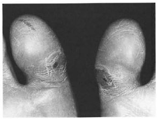
Fig. 1. Calluses with "near-healed" chronicallyrecurrent insensitive leprous ulcers.
In May and June of 1988, a total of 0.60 ml of silicone was implanted into each hallux using a tuberculin Luer-Lok syringe and a 25-gauge ⅞ inch (22-mm) needle. The fluid was injected subdermally and central to the points of maximum pressure using 0.10 ml per injection at each site over 6 weekly visits. No special shoes, shoe inserts, pads, or pressure-reducing devices were used in conjunction with the silicone. During 9 years of annual observation, neither the calluses nor the ulcers recurred or required further care (Fig. 2).
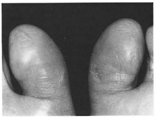
Fig. 2. Fractional silicone injections have pre vented callus and ulcer recurrence for 9 years.
Ulcer prevention in three leprosy patients (7 sites: 5 planter, 2 digital) mirrors the results comparable to those observed by Balkin and Kaplan (1) following silicone injections in insensitive diabetic feet where 23 of 29 planter ulcers injected after healing had no postinjection recurrence during 13-201 months (1-16 years), mean 6.3 years. In both leprous or diabetic patients there were no adverse reactions to the silicone, and among all patients injected, the only complication of significance was infrequent and generally asymptomatic fluid migration (1).
Over the years of this study there were many opportunities to gather biopsied as well as autopsied skin specimens for microscopic analysis. Host response to silicone fluid has been found to consist of essentially noninflammatory fibrosis and histiocytosis. The morphology exhibits a chemically and physically stable synthetic fat pad between overlying epidermis and underlying tendons, bone and joints. Countless microdroplets in and outside histiocytes are largely subdermal and are prominently distributed perineurally and perivascularly. The morphologic similarity over the years suggests a recycling of silicone into progressively smaller droplets, out from the fibrous bonds and demise of histiocytes to be followed by their return to bondage by collagen and successive generations of histiocytes. The earliest specimen studied among 124 obtained postmortem from 32 patients was 1 year and the oldest 29½ years, mean 13 years. In 11 patients permission was also granted to remove the inguinal nodes, and in four patients other lymph systems were studied as well as all major viscera. Microscopic droplets were found in the inguinal lymph nodes only, without signs or symptoms. Histologic findings suggest that injected silicone forms a safe cushion-like prosthesis that simulates fibro-fatty tissue and is remarkably well-retained long-term despite the incalculable forces which feet sustain.
In 25,000 recorded silicone injections at several thousand digital or planter pressure sites, not a single infection, fluid silicone rejection, inflammatory or allergic response has been observed. The injection technique is simple and nondisabling, and as opposed to feet with sensation does not require local anesthesia. It appears to be a most promising treatment for pressure-related disorders from which feet lacking sensitivity are at greater risk. It is entirely reasonable to assume that prevention of insensitive pressure ulcers will prevent amputations. Authorized clinical trials are warranted and if these favorable results are confirmed, the enormous disability and economic costs attending these age-old foot disorders may be dramatically reduced.
REFERENCES
1. BALKIN, S. W. and KAPLAN, L. Injectable silicone and the diabetic foot: a 25-year report. Foot 2(1991)83-88.
2. BOULTON, A. J. M, HARDISTY, C. A.. BETTS, R. P.. FRANKS, C. I.. WORTH. R. C, WARD, J. D. and DUCKWORTH, T. D. Dynamic foot pressures and other studies as diagnostic and management aids in diabetic neuropathy. Diabet. Care 6(1983)26-33.
3. BRAND. P. W. Repetitive stress in the development of diabetic foot ulcers. In: The Diabetic Fool. 4th edn. Levin, M E. and O'Neal, L. W., eds. St. Louis: C. V. Mosby. 1988, pp. 83-90.
4. CTERCTEKO, G. C, DHANENDRAN, M, HUTTON, W. C. and LE QUESNE, L. P. Vertical forces acting on the feet of diabetic patients with neuropathic ulceration. Br. J. Surg. 68(1981)608-614.
5. EDMONDS, M. E. and WATKINS, P. J. Management of the diabetic foot. In: Diabetic Neurepathy . Dyck, P. J., Thomas, P. K., Ashury, A. K., Wine-grad, A. I. and Porte. D., eds. Philadelphia: W. B. Saunders, 1987, pp. 208-215.
6. ENNA, C. D. Otbservations of the hallucal sesamoids in trauma to the denervated foot. Int.Surg. 53(1970)97-107.
7. ROSS, W. F. Etiology and treatment ol plantar ulcers. Eepr. Rev. 33(1962)25-40.
1. D.P.M. Podiatry Section, Department of Orthopaedics, Los Angeles, California, U.S.A.
2. M.D. Department of Dermatology, Los Angeles County-University of Southern California Medical Center, Los Angeles, California, U.S.A.
3. M.D. Division of Anatomic Pathology, Cedars-Sinai Medical Center, Los Angeles, California, U.S.A.