- Volume 64 , Number 3
- Page: 324–5
Penile lesion in hansen's disease
This department is for the publication of informal communications that are of interest because they are informative and stimulating, and for the discussion of controversial matters. The mandate of this Journal is to disseminate information relating to leprosy in particular and also other mycobacterial diseases. Dissident comment or interpretation on published research is of course valid, but personality attacks on individuals would seem unnecessary. Political comments, valid or not, also are unwelcome. They might result in interference with the distribution of the Journal and thus interfere with its prime purpose.
To the Editor:
Lesions on male genitals in Hansen's disease are considered to be unusual. Still rarer is the primary involvement of" preputial skin. We report one such case.
CASE REPORT
A 45-year-old male presented to us with 2 month's history of paresthesia involving the distal part of both extremities. Clinical examination revealed loss of pain, touch and temperature sensation over the dorsum of both hands and feet. Examination of the peripheral nerves showed bilaterally thickened ulnar nerves and thickened and tender right common peroneal nerve. The patient when examined for any cutaneous lesion showed one erythematous hypoesthetic plaque on the skin of the lower back. Interestingly, the skin of the prepuce also had a well-defined, shiny, erythematous plaque (Fig. 1). The lesion was extending onto the mucosal surface of the prepuce. The plaque was completely anesthetic to pinprick, touch and temperature. Cutaneous and neurological findings taken together suggested a clinical diagnosis of borderline tuberculoid Hansen's disease. A slit-skin smear was negative for acid-fast bacilli. A skin biopsy from the lesion on the prepuce showed epidermal atrophy and the presence of an epithelioid cell granuloma in the dermis with many giant cells. The histologic features were consistent with tuberculoid Hansen's disease (Fig. 2).
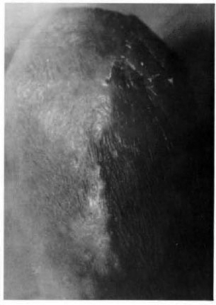
Fig 1. Anesthetic plaque on preputial skin.
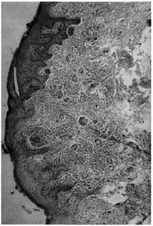
Fig. 2 Histology showing epithelioid cell granulomas in dermis with many giant cells (hematoxylin-eosin x100).
DISCUSSION
Skin lesions in Hansen's disease usually occur on the cooler regions of the body (1). The genitals are considered to be immune zones for the development of leprosy (4). However, there have been some reports on involvement of the genitalia in leprosy (2,3,5). Arora, et al. (2) in their study on 450 males with leprosy reported genital lesions in 2.9% of their patients; the lesions were seen in borderline, borderline lepromatous, and lepromatous cases. Parikh, et al. (3) described scrotal and penile lesions in six patients with borderline leprosy (BT-BL). These reports suggest that lesions on genitalia are not so uncommon as believed. However, to the best of our knowledge, lesions occurring on the prepuce involving the mucosal aspect have not been reported earlier. Under-reporting of these patients is due either to reluctance on the part of patients to expose or the physician to examine the genitalia. Our patient was unaware of his skin lesion, and the lesions were detected only after a thorough physical examination.
- Sunit Maru, M.B.B.S.
Assit Mittal, M.D.
Lalit Gupta, M.D.
Mukul Sharma, M.D.
Nirmal Bansai, M.D.
Department of DermatoVenereo-Leprology
R.N.T. Medical College
Udaipur 313 001, India
REFERENCES
1. Anish, S. A. The relationship between the surface temperature and dermal invasion in lepromatous leprosy. Int. J. Lepr. 39(1971)848-851.
2. Arora, S. K., Mukhija, R. D., Mohan, L. and Girdhar, M. A study of cutaneous lesion on male genitalia. Indian J. Lepr. 61(189)222-224.
3. Dixit. V. B., Choudhary, S. D., Jain, V. K. and Rajeev, S. Primary involvement of scrotum in tuberculoid leprosy. Indian J. Lepr. 62(1990)120-121.
4. Fox, H. and Knott, J. Leprous nodules of male genitalia. Int. J. Lepr. 2(1934)445-446.
5. Parikh, D. A., Parikh, A. C. and Ganarati. R. Penile and scrotal lesions in leprosy: case report. Lepr. Rev. 60(1989)303-305.
Reprint requests to Dr. A. Mittal, 3 Seth Ji Ki Bari, Madhuvan, Udaipur 313 001, India.