- Volume 63 , Number 3
- Page: 454–5
Tuberculous optic neuritis histologically resembling leprous neuritis
To the Editor:
We wish to report an interesting histopathological observation in the optic nerve of a case of ocular tuberculosis. A 5-year-old female underwent enucleation of the blind left eyeball for suspected retinoblastoma following detection of a pupillary white reflex, shallow anterior chamber, iris neovascularization and secondary glaucoma. On clinical examination right axillary lymphadenitis was found. This was diagnosed to be tuberculous on fine-needle aspiration cytology.
Histological examination of the eyeball revealed widespread tuberculosis involving uveal tissue and the retina. A section through the resected end of the optic nerve (The Figure) showed epithelioid cell granulomas in the substance of the nerve, reminiscent of neuritic leprosy. Tuberculous infection of the optic nerve is rare. It spreads from tuberculous lesions in the eye, brain or the adjacent orbital structures (1,2).
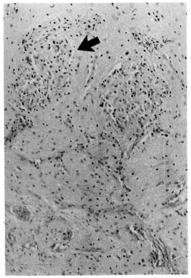
The figure. Granulomas (arrow) in the optic nerve.The optic nerve is distinguished by its glial appearance in histological sections (hematoxylin and eosin x 100).
Leprosy, on the other hand, is a disease of the peripheral nerves. Demonstration of Mycobacterium leprae or granulomas in nerves arc the mainstay of histopathological diagnosis. Granulomas in a nerve have the same diagnostic significance as the presence of M. leprae (8). This is explained by the fact that M. leprae are the only known neurotropic mycobacteria (4). Cranial nerves with a peripheral component, i.e., the facial and the trigeminal, are known to be affected in leprosy. The optic nerve escapes probably because it is devoid of Schwann cells (3).
Other mycobacteria, not being neurotropic, do not primarily cause neuritis. However, granulomatous inflammation is destructive of pre-existing tissue (7), and nerves caught up in the inflammatory process may be involved. This fact probably explains the sporadic case reports of evidence of nerve involvement in nonleprosy granulomatous dermatoses (5,6)- with accompanying sensory impairment.
We have reported earlier our observations of different patterns of dermal nerve involvement in lupus vulgaris, granulomatous secondary syphilis and other non-leprosy granulomas (9). A granuloma within a dermal nerve was found in a case of lupus vulgaris. The present case documents granulomatous involvement of a large cranial nerve with tuberculosis.
- Vinod K. Arora, M.D.
Reader, Department of Pathology
- Upreet Dhaliwal, M.S.
Lecturer, Department of Ophthalmology
- Navjeevan Singh, M.D.
Reader, Department of Pathology
- Arati Bhatia, M.D.
Professor, Department of Pathology
University College of Medical Sciences and Guru Tegh Bahadur Hospital
Shahdara, Delhi 110 095, India
Acknowledgment. The authors wish to thank Mr. Har Prasad and Mr. Ashish Oberoi of the UCMS photo unit for their assistance.
REFERENCES
1. Cook, C. A. G. and Morgan, G. The eyes. In: Systemic Pathology. 2nd edn. Vol. 6. Symmers, W. St.C, cd. Edinburgh: Churchill Livingstone, 1980, pp. 2833-2862.
2. Duke-Elder, S. and Scott, G. I. The optic nerve: specific infections - tuberculosis. In: System of Ophthalmology. Vol. 12. London: Henry Kimpton, 1971, pp. 167-173.
3. Ham , A. W. and Leeson, T. S. The cells of the nervous tissue of the central nervous system. In: Histology. 4th. edn. London: Pitman Medical Publishing Co., Ltd., 1961, pp. 465-466.
4. Iyer, C. G. S. prediliction of Mycobacterium leprae for nerves. Int. J. Lepr. 33(1965)634-645.
5. Kubba, R., EL-Hassan, A. M., AL-Gindan, Y., Omer, A. H., Busra, M. and Kutty, M. K. Peripheral nerve involvement in cutaneous leishmaniasis (Old World). Int. J. Dermatol. 26(1987)527-531.
6. Mann, R. J. and Harman, R. M. Cutaneous anesthesia in necrobiosis lipoidica. Br. J. Dermatol. 110(1984)323-325.