- Volume 63 , Number 1
- Page: 28–34
Optimal pH for preserving the activity of Mycobacterium leprae during Incubation of cells in a cell-free liquid medium
ABSTRACT
The effect of the pH of a cell-free liquid medium on the activity of Mycobacterium leprae during incubation of the cells was investigated. As a parameter for evaluating the activity, the amount of adenosine triphosphate (ATP) extracted F rom the incubated cells collected by ccntrifugation was measured. The results demonstrate that the activity of M. leprae cells was maintained at a significant level for approximately 4 weeks at 30ºC in 0.05 M phosphate buffer containing 10% fetal calf serum at pH 7.0 compared to cells at other pHs tested, but activity was not preserved in phosphate buffer at pH 7.0 without serum and incubated at 37ºC. The maintenance of the activity under these conditions was prolonged somewhat by the addition of glycerin (2%) to the medium, and was definitely inhibited by rifampin but not by either penicillin or isoniazid. F rom the results reported here, it could be postulated that the optimal pH of cell-free media for the study of cultivation of M. leprae is 7.0.RÉSUMÉ
On a examiné l'effet du pH d'un milieu liquide acellulairc sur l'activité de Mycobacterium leprae durant l'incubation des cellules. Comme paramètre d'évaluation de l'activité, on a mesuré la quantité d'adénosine triphosphate (ATP) récoltée par ccntrifugation à partir des cellules incubées. Les résultats démontrent que l'activité des cellules de M. leprae était maintenue à un niveau significatif pour environ 4 semaines à 30ºC dans un tampon phosphate 0.05 M contenant 10% de serum de foetus de veau à pH 7.0, par comparaison à des cellules testées à d'autres pH, mais l'activité n'était pas préservée dans un tampon phosphate à pH 7.0 sans serum et incubée à 37ºC. La maintenance de l'activité dans ces conditions était quelque peu prolongée par l'addition de glycérine (2%) au milieu, et était nettement inhibée par la rifampicine, mais non par la pénicilline ni par l'isoniazide. A partir de ces résultats, on pourrait postuler que le pH optimal des milieux acellulaircs pour l'étude de la culture de M. leprae est 7.0.RESUMEN
Se investigo el efecto del pH de un medio libre de células sobre la actividad metabólica de Mycobacterium leprae incubado en ese medio. Como parámetro de la actividad se midió la cantidad de trifosfato de adenosina (ATP) extraído de las células colectadas por centrifugación. Los resultados demostraron que la actividad de M. leprae se mantuvo a niveles significantes a lo largo de 4 semanas de incubación a 30ºC en regulador de fosfatos 0.05 M, pH 7.0, con 10% de suero fetal de ternera. La actividad del microorganismo no se preservó cuando la incubación se hizo a 37ºC en regulador de fosfatos, pH 7.0, sin suero fetal de ternera. Bajo estas últimas condiciones la actividad se prolongó un poco cuando se adicionó glicerol al 2% al medio, y se inhibió completamente por la rifampina pero no por la penicilina o la isoniazida. De estos resultados se peude postular que el pH óptimo para el cultivo de M. leprae en medios libres de células es de 7.0.Since the discovery of Mycobacterium lepraemurium, numerous attempts to cultivate the mycobacterium in vitro have been made for about 70 years. In 1969, however, Ogawa and Motomura (13) succeeded in inducing continuous colony formation of the bacilli on solid egg-yolk medium. The results were confirmed by Mori (10) and Pattyn, et al. (14). Unequivocal multiplication of the bacilli in a cell-free liquid medium was independently achieved by Nakamura (11). This finding was confirmed by Dhoplc and Hanks (3) using parameters of bacillary counting and measurement of ATP.
The key to these achievements in the cultivation and multiplication of M. lepraemurium was only that the pH of the culture medium should be adjusted to 6.0. When the pH of the medium was adjusted to 6.0, M. lepraemurium started to grow on/in the medium which is routinely used for M. tuberculosis, although its growth was less than that obtained on/in the media developed by Ogawa and Motomura as well as Nakamura. To adjust the pH of the medium to 6.0, Na2 HPO4 is omitted from the original solid Ogawa medium and α-ketoglutaric acid is added to the Kirchncr liquid medium (7). Therefore, it is absolutely critical for the growth of M. lepraemurium that the pH of the medium be 6.0.
M. leprae, the causative agent of leprosy, has still not been cultivated in vitro, either in a tissue culture or in a cell-free medium, even more than 100 years after the discovery of the bacillus. It is conceivable, from the successful cultivation of M. lepraemurium, that the pH of the culture medium might be one of the critical factors affecting the multiplication of M. leprae also.
The present paper reports the optimal pH for preserving the activity of M. leprae in cell-free liquid media during incubation of the bacilli.
MATERIALS AND METHODS
Preparation of M. leprae suspension. An M. leprae suspension was prepared as previously described (12). Briefly, foot pads of nulnu mice experimentally infected 12 months previously with 1 x 10⁶ M. leprae Thai 53 strain (9) (kindly supplied by Dr. M. Matsuoka, National Institute for Leprosy Research, Tokyo) were surface decontaminated with iodine tincture for 1 min, then rinsed in 70% ethanol. The foot pads were then minced with scissors and homogenized with a glass homogenizer in sterile 0.05 M phosphate buffer (pH 7.0). After the coarse tissue debris was removed by lowspeed ccntrifugation (100 x g x 3 min), sterile 0.5% w/v trypsin (Sigma Chemical Co., St. Louis, Missouri, U.S.A.) solution was added to the bacillary suspension at a final concentration of 0.05%. The mixture was incubated in a water bath at 37ºC for 45 min, then centrifuged (1500 x g x 15 min) to remove the trypsin. The sediment was suspended in 0.05 M buffer (pH 7.0) and treated with NaOH at a final concentration of 0.25 N for 15 min in a water bath at 37ºC. After this treatment, the mixture was centrifuged at 1500 x g x 10 min. The final suspension was obtained by resuspending the pellet in 0.05 M phosphate buffer (pH 7.0) containing the fetal calf serum (10% v/v).
No reduction of the activity compared to that of the starting material was found by inoculation into mouse foot pads.
Preparation of M. lepraemurium suspension. A leproma produced in a C3H mouse experimentally inoculated subcutaneously with the Hawaiian strain (M-85) M. lepraemurium was homogenized in 0.05 M phosphatc bufFer (pH 6.5) in a glass homogenizer, then treated with NaOH (final concentration, 0.25 N) for 15 min in a water bath at 37ºC. Then the mixture was ccntrifuged at 1500 x g x 10 min. The final suspension was made by resuspending the pellet in a phosphate buffer containing fetal calf scrum (10%v/v).
Inactivation of M. leprae. For inactivating the cells by ultraviolet (UV) irradiation, 10 ml of a bacillary suspension was transferred to an open Petri dish (10 cm in diameter), and exposed at a distance of 15 cm to a 10-watt germicidal lamp (National Electric Co., Japan) for 30 min. During irradiation, the pctri dish was agitated alternately in a vertical and then in a horizontal direction. For inactivation by heat, 5 ml of the M. leprae suspension in a screw-cap tube was heated at 120ºC for 10 min. No tests for the remaining infectivity of M. leprae after treatments were carried out.
Culture medium. Phosphate buffers (KH2PO4:Na2HPO4) (0.05 M) of different pHs were used as basal culture media. Changes in the pH caused by addition of fetal calf serum (GIBCO BRL, Gaithersburg, Maryland, U.S.A.) at a final concentration of 10% to the sterile buffers were as follows: the original pH 5.5 to 5.75-5.78, 6.0 to 5.95-5.96, 6.5 to 6.44-6.45, 7.0 to 6.95-6.96, and 7.5 to 7.42-7.44. [In the text, the pH of the culture medium before the addition of the serum, not the pH after the addition of serum, is designated as the original pH. The phosphate bufTer containing serum is referred to as serum-buffer.]
Inoculation of bacillary suspension and incubation conditions. Six-tenths ml of a suspension containing 3.3-5.6 x 10⁸ M. leprae was inoculated into 40 ml of scrumbuffer. In the case of M. lepraemurium, 1 x 10⁷ cells were used. The culture tubes (90 mm x 12 mm, screw-cap tubes) contained 5.5 ml of the inoculated medium, leaving a 27.7% free air space above the medium. Inoculated culture tubes generally were incubated at 30ºC.
Chemotherapeutic agents. Penicillin G (Tomiyama Chemical Industries, Japan), isoniazid (Dai-ichi Chemical Industry Co., Japan) and rifampin (CIBA-GEIGY Ltd, Japan) were used.
ATP extraction. ATP determinations were carried out by the firefly bioluminesccnt technique (2-6). Tubes of bacterial suspension at zero-time and those incubated for various periods of time were centrifuged for 20 min at 3000 x g, then 4.5 ml of the supernatant was discarded and the remaining suspension was vortexed briefly. These suspensions, approximately 1 ml, were completely transferred to disposable 1.5-ml Eppendorf tubes which were centrifuged at 12,500 x g x 6 min to collect the cells. The supernatant was discarded, and the pellet was suspended in 0.1 ml of Tris-1 mM EDTA pH 7.7 and vortexed briefly. Thereafter, 0.2 ml of 100 mM Tris containing 2 mM EDTA pH 7.75 was added, and the suspension was vortexed again. Chloroform (0.04 ml) was added to the suspension, and the contents were mixed well. The tube was placed in a boiling water bath for 7 min. After the mixture was cooled, 0.9 ml of distilled water was added to make a test sample. Onc-tcnth ml of lucifcrin-luciferase was injected into 0.4-ml portions of the test samples. An Aminco Integrator (American Instrument Co., Silver Spring, Maryland, U.S.A.) was used to determine ATP values. The actual amounts of ATP per tube could be calculated by multiplying the measured values by three.
To describe the stability of ATP extracted from M. leprae cells, all values were expressed as a percent of the original suspension at zero-time.
Bacteriological examinations. The M. leprae used in all inocula, as well as those from all in vitro cultures harvested, and contamination checks were characterized on the basis of the following criteria: acid-fastness by Zichl-Ncclscn staining, inability to grow on blood agar and Ogawa egg slants, and pyridine extraction of acid-fastness of stained bacilli.
Morphological features of the bacilli in the inoculum and in tubes incubated for various periods were observed after Ziehl-Neclsen staining, and the bacilli were counted by the method of Shepard and McRac (15).
RESULTS
Effect of pH of serum-buffer. It may be emphasized that, from the results obtained from more than five separate experiments, the presence of more than 2000 pg of ATP extracted from the cells of M. leprae per tube in the starting sample was essential for obtaining the reproducible results reported below.
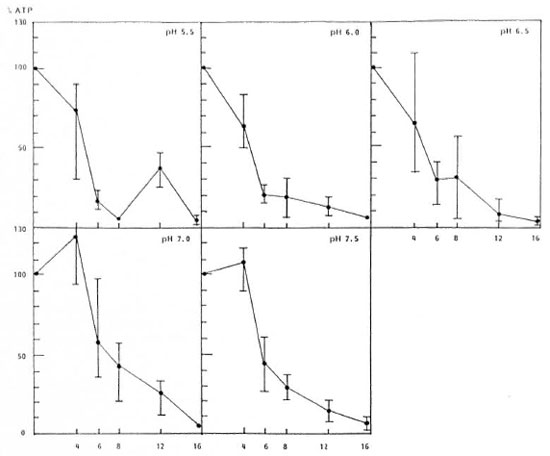
ATPs were extracted from the cells at periodic intervals during cell incubation in scrum-buffers of different pHs. The results shown in Figure 1 indicate that the yields of ATP extracted from the cells gradually decreased with time of incubation at 30ºC in serum-buffer at pH 5.5, 6.0 and 6.6. In serum-buffer at pH 7.0 and 7.5, however, approximately 30%-50% and 10% increases in ATP over the original zero-time sample, respectively, were observed after 4 weeks of incubation, and then decreased.

Fig. 1. Stability of At leprae during incubation of cells in serum-buffers of different pHs at 30ºC.
No differences between the declining slopes of the ATP yields with incubation time were seen in the serum-buffers of different pHs, except pH 5.5 at which a slight increase in ATP was observed at 12 weeks of incubation compared to the values at 8 weeks.
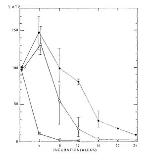
Effects of serum and glycerin. As shown in Figure 2, when fetal calf serum was omitted from the serum-bufTer, i.e., a simple phosphate buffer pH 7.0 alone, the activity of M. leprae in the buffer was rapidly lost within 2 weeks of incubation. Ifglycerin was added to the serum-buffer at the final concentration of 2% (v/v), the ATP yield was slightly increased during 4 weeks of incubation and then declined. The rate of decline was slower than that in the original serum-buffer.

Fig. 2. Stability of M. leprae during incubation of cells in serum-buffer (0.05 M, pH 7.0) with ( ), and without (
 ) glycerin, and without fetal calf serum (
 ).
).
Effect of incubation temperature. When M. leprae cells were incubated in serumbuffer pH 7.0, at 37ºC, no sustainmcnt or increase in the amount of ATP extracted from the incubated cells was demonstrated: 30% to 70%, and 10% to 30% of the amount of ATP extracted from the zero-time sample were extracted after 4 and 8 weeks' incubation, respectively.
ATP yields from inactivated cells. Approximately 70%-90% of the amount of ATP extracted from the starting sample was extracted from UV-killed cells of M. leprae just after irradiation. When the UV-killed cells were incubated in the serum-bufFer pH 7, no ATP was extracted from the cells after 2 weeks of incubation.
No ATP was extracted from the heatkilled cells of M. leprae just after heating.
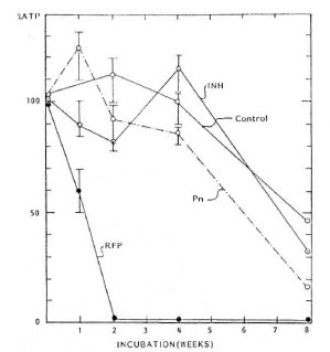
Effect of chemotherapeutic agents. The effects of penicillin (final concentration 100 Mg/ml), isoniazid (10 Mg/ml) and rifampin (20 Atg/ml) on the ATP yields extracted from incubated M. leprae cells were investigated. The results are shown in Figure 3. Only rifampin inhibited the maintenance of the ATP activity of M. leprae.

Fig. 3. Effects of penicillin (Pn; 100 µg/ml), isoniazid (INH; 10 µg/ml) and rifampin (RFP; 20 µg/ml) on the stability of M. leprae during incubation of cells in the serum-buffer.
Morphological observations. No significant increase in the number of cells during incubation was seen. In some samples, there was an increase in the number of cells but it could not be distinguished from the apparent increase in cells resulting from declumping, in which large aggregates of M. leprae in the inoculum are dispersed during incubation. In addition, no significant difference in cell length was observed between the cells at zero-time and cells harvested after the various periods of incubation.
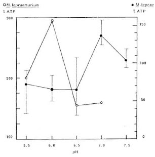
Optimal pH for ATP concentration of M. lepraemurium. It is known that growth of M. lepraemurium takes place when the cells are inoculated into the established culture medium with the pH adjusted to 6.0. Yields of ATP extracted from the cells of M. lepraemurium incubated for 4 weeks in the serum-buflcrs having different pHs were measured. The results indicate, as shown in Figure 4, that the largest amount of ATP could be extracted from those cells that were incubated in the serum-bufTer at pH 6.0.

Fig. 4. Stability of M. leprae ( ) and M. lepraemarium (
 ) during incubation at 4 weeks and 8 weeks, respectively, of cells in serum-buffers at different pHs.
DISCUSSION
A definite amount of M. leprae ATP in the starting material for obtaining reproducible results described here was found to be necessary. Although the reason for this requirement is not clear, the activity of M. leprae in a cell-free liquid medium could be maintained at a level at least as high as at zero-time, and even somewhat higher for approximately 4 weeks at 30ºC, when the cells of M. leprae were incubated in 0.05 M phosphate buffer pH 7 containing 10% fetal calf scrum. The activity of the cells was evaluated by measuring the amount of ATP extracted from the incubated cells.
There are three possible factors influencing the yield of ATP extracted from the incubated cells because the ATP was extracted from the cells collected by ccntrifugation: a) The amount of ATP represents the sum of the ATP produced by genuine energy metabolism initiated in a serum-buffer at pH 7 when the cells were incubated at 30ºC. b) The yield of ATP indicates an increase in the total weight of individual cells during incubation, caused by an increase in the number of cells collected by ccntrifugation. c) The increase in the amount of ATP could be due to a decrease in viscosity of the culture medium as a result of chemical denaturation during incubation in which the cells are more rapidly sedimcntcd and more efficiently harvested than in the starting medium.
Among these three possibilities, the results obtained strongly suggest that the yield of ATP extracted from the incubated cells represents the total amount of ATP generated and accumulated in continuation of the energy metabolism of the M. leprae cells in the serum-buffer at pH 7, during incubation at 30ºC. This is because the maintenance of the activity occurred only in the medium containing fetal calf scrum within the limited pH range of 7 at 30ºC but not at 37ºC. In addition, the maintenance of the activity of the cells was prolonged by adding glycerin to the medium; it is well known that glycerin is important as a carbon source for mycobacteria. Therefore, it might be implied that the M. leprae cells have utilized serum and glycerin as an energy source. In other words, it is obvious that the amount of ATP extracted from the incubated cells definitely depends on several factors, such as serum and glycerin, as well as incubation temperature.
The ATP extracted from the cells after 4 weeks' incubation is not likely the initial ATP present in the starting cells but ATP produced during incubation, because no ATP was obtained from the UV-irradiatcd cells without metabolism after 2 weeks' incubation; whereas 70%-90% of the ATP of the intact cells could be obtained from the UV-killed M. leprae just after irradiation. ATP in the cells without metabolism might diminish rapidly during incubation. Furthermore, the maintenance of the activity under the limited condition was definitely inhibited by rifampin but not by either penicillin or isoniazid. These findings are quite similar to those for the response of M. leprae to chcmothcrapcutic agents reported by Franzblau and Hastings (5).
Finally, the reason why pH 7.0 should be considered optimal for the growth of M. leprae is the fact that the increase in ATP extracted from the cells of M. lepraeniuriuni was observed when the cells were incubated in the serum-buffer at pH 6. It is evident that the pH 6.0 of the established solid medium as well as liquid medium is surely optimal to support the growth of M. lepraeniuriuni.
These results might conflict with the data reported by others. The discrepancy between the results reported by Lee and Colston (8), indicating a rapid decay in intracellular ATP of M. leprae under the cell-free condition, and the results of the experiment described here might be due to the difference in the kind of culture medium and its pH.
On the other hand, the data demonstrating a slow decay of intracellular ATP of M. leprae in Dubos albumin medium, pH 7.0 (5), as well as in Dhoplc-Hanks medium, pH 7.2 (1), might support the results reported here, although the compositions of the culture media used were different.
It cannot be concluded whether the slight increase in ATP extracted from the cells incubated in the scrum-buffer pH 5.5, after an incubation of 12 weeks, is due to ATP extracted from the cells adapted from the in vivo to the in vitro environment, as described by Dhople and Hanks (3), in the experiment on M. lepraeniuriuni, because the culture medium used here is a nutrientpoor solution, i.e., serum-buffer, rather than a nutrient-rich medium.
It is well known that some bacteria require a strictly limited pH of the appropriate medium to support growth; for instance, Vibrio cholerae requires peptone water of pH 8, Legionella pneumophila requires a yeast extract-cysteine medium of pH 6.95 ± 0.05 (4), and the growth of M. lepraemurium is observed only in/on a medium of pH 6.0, as previously mentioned. Therefore, it is easy to presume that M. leprae also might require a limited pH of the culture medium, i.e., pH 7, to support the energy metabolism necessary for multiplication.
Acknowledgment. I am grateful to Prof. Y. Hagihara, Dean of the Fukuoka Dental College, for his generous consideration in allowing me to use the apparatus for measuring ATP.
REFERENCES
1. DHOPLE, A. M., GREEN, K. J. and OSBORNE, L. J. Limited in vitro multiplication of Mycobacterium leprae. Ann. Inst. Pasteur/Microbiol. 139(1988)213-223.
2. DHOPLE, A. M. and HANKS, J. H. Quantitative extraction of ATP from cultivable and host grown microbes: calculation of ATP pools. Appl. Microbiol. 26(1973)399-403.
3. DHOPLE, A. M. and HANKS, J. H. Factors that influence the growth of Mycobacterium lepracmurium in the Nakamura system. Int. J. Lepr. 44(1976)18-26.
4. FEELEY, J. C, GORMAN, G. W., WEAVER, R. E., MACKEL, D. C. and SMITH, H. W. Primary isolation media for Legionnaires disease bacterium. J. Clin. Microbiol. 8(1978)320-325.
5. FRANZBLAU, S. G. and HASTINGS, R. C. Rapid in vitro metabolic screen for antileprosy compounds. Antimicrob. Agents Chemother. 31(1987)780-783.
6. KATOCH, V. M., KATOCH, K., BHARADWAJ, V. P., DATTA, A. K., SHARMA, V. D., SHIVANNAVAR, C. T. and KANNA, K. B. Metabolic studies on mycobacteria. V. A preliminary report on the ATP synthesis by mycobacteria including M. leprae by using different substrates. Indian J. Lepr. 59(1987)163-170.
7. KIRCHNER, O. Die Leistungsfähigkeit der Tiefcnkultur des Tuberkclbazillus bei Verwendung besonders geeigneter flüssiger Nährboden. Zentl. Baktcriol. Parasit. Infek. Hyg. 124(1932)403-412.
8. LEE, Y. N. and COLSTON, M. J. Measurement of ATP generation and decay in Mycobacterium leprae in vitro. J. Gen. Microbiol. 131(1985)3331-3337.
9. MATSUOKA, M., KOHSAKA, K. and DAYANGHIRANG, J. A. Characterization of Mycobacterium leprae Thai-53 strain.(Abstract)Int. J. Lepr. 60(1992)722.
10. MORI, T. Study of a growth factor for Mycobacterium lepraemurium 1. Minimal medium. Int. J. Lepr. 46(1978)125-132.
11. NAKAMURA, M. Multiplication of Mycobacterium lepraemurium in cell-free medium containing a-kctoglutaric acid and cytochrome c. J. Gen. Microbiol. 73(1972)193-195.
12. NAKAMURA, M. Elimination of contaminants in a homogenate of nudc-mousc footpad experimentally infected with Mycobacterium leprae. Jpn. J. Lepr. 1994(in press).
13. OGAWA, T. and MOTOMURA, K. Studies of Mycobacterium lepraemurium I. Attempts to cultivate M. lepraemurium. Lepro 38(1969)246-254.
14. PATTYN, S. R. and PORTAELS, F. In vitro cultivation and characterization of Mycobacterium lepraemurium. Int. J. Lepr. 48(1980)7-14.
15. SHEPARD, C. C. and MCRAE, D. H. A method for counting acid-fast bacteria. Int. J. Lepr. 36(1968)78-82.
M.D., Ph.D., Professor Emeritus, Kurume University, Koga Hospital Medical Research Institute, 120 Tenjin, Kurume 830, Japan.
Received for publication on 26 July 1994.
Accepted for publication in revised form on 29 September 1994.