- Volume 63 , Number 1
- Page: 101–3
Mycobacterium leprae in the epidermis: ultrastructural study I
To the Editor:
Mycobacterium leprae is an intracellular bacterium which is located mainly in the dermal and subcutaneous regions of the skin. In the skin lesion, there is known to be a clear zone separating the epidermis from the granuloma in both borderline lepromatous (BL) and lepromatous (LL) leprosy. In our present study, we examined the skin biopsy sample from a male BL patient, 51 years old, under the electron microscope. The sample was prepared with routine procedures for electron microscopy. Ultrathin sections of the specimen were stained in saturated uranyl acetate and lead citrate separately before they were examined in a Phillips CM10.
The samples revealed some bacilli in the epidermal cells. Just above the basement membrane of the epidermis, there were several cells containing M. leprae in their cytoplasm without any membrane structure around the bacilli (Fig. 1). These bacilli looked free in the cytoplasm of the epidermal cells (Fig.2). The cells in which the bacilli are located are the typical epidermal cells, having tonofilaments (T) and melanosomes (M) in their cytoplasm and also desmosomes (D) at the junction of each of the cells. Beneath the basement membrane, there is a cell filled with the bacilli (Fig.3). This cell has well developed dendrites on its surface.
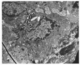
Fig. 1. M. leprae (B) in epidermal cells. T = tonofilament; M = melanosome; D = desmosome: BM =basement membrane. (Bar = 2 µm)
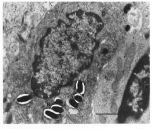
Fig. 2. Higher magnification of a keratinocyte. (Bar = 1 µm)
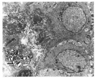
Fig. 3. M. leprae in a dendritic cell in the dermal region. DC = dendritic cell. (Bar = 2 µm)
There have been reports (2,6) about the epidermal localization of M. leprae in lepromatous leprosy. In their report, Okada, et al .(6) suggested that the bacilli can be phagocytized by the keratinocyte, but they did not mention where the bacilli meet the keratinocyte.
There are several kinds of dendritic cells in the dermis, such as melanocytes, Lan- gerhans cells, and Merkel cells. These cells can move from the dermis to the epidermis and among these cells, Langerhans cells can move up and down through the basement membrane. These cells also have some phagocytic abilities. According to the reports from Cramer (1) and Hulley, et al. (3), melanocytes migrate up from the dermis into the epidermis, not only in normal development, but also during normal tissue maintenance. Also, Klaus (4) showed the transfer of melanosomes from melanocytes to keratinocytes. With all of these previous reports we suggest that the dendritic cell(s) in the dermis can take up (or get) the bacilli and transfer them to the keratinocyte by the same method as that used in the transfer of melanosomes from melanocytes to keratinocytes (4,5).
The possibility that the dendritic cell can transfer the bacilli to the epidermis and the identity of the dendritic cell need more verification. We also found a bacillus in a Langerhans cell (unpublished data) of a LL patient's epidermis.
With the progress of keratinization, the bacilli will go up to the keratin layer and eventually be removed from the skin. The question is whether the bacilli are alive or not when they leave the skin. The significance of the existence of M. leprae in the epidermis is not as yet explained, but more information about the epidermal bacilli should give us the answer.
Finally, this study suggested several possibilities in the progress of leprosy: 1) A dendritic cell can transfer the bacilli to the epidermis although the mechanism of uptake of the bacilli by the dendritic cell and the keratinocytcs is not known. 2) The bacilli can be discharged from the skin of the patients, although the condition of the bacilli is not yet known.
- Young-Hoon Seo, Ph.D.
- Wook Cho, M.D.
- Hae-Young Choi, M.D.
- Yong-Ma Hah, M.D., Ph.D.
Institute for Leprosy Research
Korean Leprosy Control Association
Anyang, Korea
- Sang-Nae Cho, D.V.M., Ph.D.
Department of Microbiology
College of Medicine
Yonsei University
Seoul, Korea
Acknowledgment. This work was supported by the Institute for Leprosy Research, Korean Leprosy Control Association. Our special thanks to Mrs. Kyung Hee Hwang and Mr. Dong Yong Chung for their excellent technical support in the electron microscopy work.
REFERENCES
1. CRAMER, S. F. The origin of epidermal melanocytes; implications for the histogenesis of nevi and melanomas. Arch. Pathol. Lab. Med. 115(1991)115-119.
2. HARADA, K. A modified alochrome procedure for demonstrating mycobacteria in tissue sections. Int. J. Lepr. 45(1997)49-51.
3. HULLEY, P. A., STANDER, C. S. and KIDSON, S. H. Terminal migration and early differentiation of melanocytes in embryonic chick skin. Dev. Biol. 145(1991)182-184.
4. KLAUS, S. N. Pigment transfer in mammalian epidermis. Arch. Dermatol. 100(1969)756-762.
5. MOTTAZ, J. H. and ZELICKSON, A. S. Melanin transfer: a possible phagocytic process. J. Invest. Dermatol. 49(1967)605-610.
6. OKADA, S., KOMURA, J. and NISHIURA, M. Mycobacterium leprae found in epidermal cells by electron microscopy. Int. J. Lepr. 46(1978)30-34.
Reprints requests to Dr. Young Hoon Seo, The Institute for Leprosy Research, Korean Leprosy Control Association, Anyang P.O. Box 27, Kyunggi-do 430-600, Korea.