- Volume 63 , Number 1
- Page: 103
Detection of AFB in tuberculoid biopsies
To the Editor:
The diagnosis of tuberculoid leprosy is made with a good margin of security since the characteristic clinical findings are associated with histological features of tuberculoid granulomatous reaction with involvement and fragmentation of the dermal nerves. The diagnosis becomes more certain and definitive if Mycobacterium leprae are detected, usually in dermal nerves or in the remains of nerve fibers. In this sense, the frequency of detection of acid-fast bacilli (AFB) in biopsies of patients with tuberculoid leprosy is underestimated. Indeed, there is a consensus concerning the rarity of AFB in these cases, figures not exceeding 7% of cases(2).
In a review of the archives from the Department of Pathology of the Instituto Lauro de Souza Lima (Bauru, Brazil) between 1980 and 1992, we came across the following data:
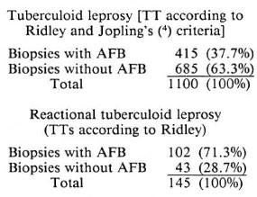
We used Faraco-Fite staining (1,3) and on each slide we put the largest possible number of sections. The section close the slide edge with the identification label is exhaustively examined. At the same time, dermal nerves and the remains of nerve fibers, if present, are localized. If AFB are found in this section, the search is concluded. If not, AFB are searched for exhaustively in the dermal nerves and/or fragments. If we increase the number of slides to be examined, the frequency of AFB found also increases, although this procedure is not viable as a routine.
- Raul N. Fleury, M.D., Ph.D.
Assistant Professor of Pathology
University of São Paulo/Bauru
Director, Department of Pathology
- Cristina M. Aranda, M.D.
Dermatologist
Instituto Lauro de Souza Lima
P. O. Box 62
Bauru, SP, Brazil 17.0001-970
REFERENCES
1. FITE, G. L., CAMBRE, P. J. and TURNER, M. H. Procedure for demonstrating lepra bacilli in paraffin sections. Arch. Pathol. 43(1947)624-625.
2. LEVER, W. F. and SCHAUMBURG-LEVER, G. Hislopathology of the Skin. 7th edn. Philadelphia: J. B. Lippincott Co., 1990, p. 256.
3. RIDLEY, D. S. Skin Biopsy in Leprosy. Basle: CIBA-GEIGY, 1984, pp. 14, 15, 42.
4. RIDLEY, D. S. and JOPLING, W. H. Classification of leprosy according to immunity; a five-group system. Int. J. Lepr. 34(1966)255-273.