- Volume 63 , Number 1
- Page: 104–5
Polyaxonal myelination in a human leprous nerve
To the Editor:
The presence of aberrantly myelinated fibers was reported by us earlier in the sciatic nerves of mice inoculated in the foot pad with Mycobacterium leprae (3) . Subsequently it was noted that mice treated with high concentrations of dapsone (DDS) or with immunomodulating regimens, such as thymectomy-irradiation or cyclosporin A, when followed by M. leprae infection showed a higher incidence of polyaxonal myelination (4,5). Such aberrant myelination also has been reported in the developing sciatic nerves of mice with benign muscular dystrophy(1,2). However, so far we have not come across any documented evidence for the presence of similar polyaxonal myelination in human nerves.
In this communication we wish to record the presence of polyaxonal myelination in a human leprous nerve. This was observed in a sural nerve biopsy obtained from a pure neuritic case of leprosy. This 17-year-old male had involvement of multiple nerves, the left lateral popliteal (common peroneal), left sural and left median nerve were thickened, The left sural nerve funicular biopsy was obtained 1 year after the MDT-MB WHO regimen(6). The nerve biopsy was studied using both light- and electron-microscopy and revealed an active borderline (BB-BL) lesion. The bacterial index in the nerve was nearly 4+ while multiple skin smears were negative throughout. Most of the bacterial load was seen within the Schwann cells (Fig. 1). There was an extensive loss of myelinated fibers. The endoncurium was well populated with Schwann cells, with and without any axonal sprouts. Occassional, small-size, thinly myelinated fibers were seen scattered all over the funicle. Dense perivascular foci of lymphocytes were present both within the endoneurium and in the epineurium. A few fibroblast-like cells with elongated processes were seen encircling groups of Schwann cells and axons. The aberrantly myelinated fiber seen in this nerve was large in size. Several axons and Schwann cell processes were seen encircled by a comparatively thin ring of myelin with normal periodicity (Fig. 2). Inner and outer mesaxons were clearly seen. The Schwann cell ensheathing this group of fibers appeared normal.
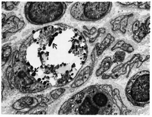
Fig. 1. Transverse section of sural nerve. A low-power electron micrograph showing leprae (arrows) in a Schwann cell of an unmyelinated fiber. Note in-crease in number of Schwann nuclei (N) ( x4500).
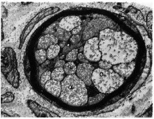
Fig. 2. Another arca in Fig. 1 nerve showing groupsof axons (a) and Schwann processes (s) surrounded bya thin ring of myclin with normal periodicity. Innerand outer mesaxons are cicarly seen ( x 12,000).
Although rare, this finding shows that polyaxonal myelination similar to that seen In mice also may occur in human leprosy lesions. The exact pathogenetic mechanism involved in such aberrant myelination is not clear. We have speculated that the occurrence of myelination around multiple axons in the mouse leprosy model is a misguided regenerative response following partial denervation of unmyelinated fiber groups-a result of defective Schwann cell axon interaction(4). As in the mouse studies, the rarity of this finding in human leprosy also could be attributed to two possible factors. Firstly, such abnormal regeneration occurred only during the early stages of infection, i.e., between the fourth and eighth postinoculation months. Secondly, a detailed study of both longitudinal and transverse sections of these nerves in mice revealed that such myelination occurred along a smail segment of otherwise unmyelinated fibers, and the axons within the myelin ring system subsequently fused to give an appearance of a single axon. According to Brown and Radich(1) such fibers undergo degeneration, thus suggesting that it is a transient phenomenon. It is not surprising therefore that such fibers are only rarely seen in human leprosy lesions.
- Dr. Vanaja Shetty
Senior Research Officer
The Foundation for Medical Research
84-A R.G. Thadani Marg, Worli
Bombay 400018, India
REFERENCES
1. BROWN, M.J. and RADICH, S.J. Polyaxonal myelination in developing dystrophic and normal mouse nerves. Muscle Nerve 2(1979)217-220.
2. OKADA, E., MIZUHIRA, V. and NAKAMURA, H. Abnormally combined myelinated and unmyelinated nerves in dystrophic mice. J. Neurol. Sci. 33(1977)243-249.
3. SHETTY, V,F. and ANTIA, N.H . Myelination around multiple axons in peripheral nerves; an unusual ultrastructural observation. Acta Neuropathol. 50(1980)147-151.
4. SHETTY, V.P. and ANTIA. N.H. Multiple axonal myelination in the experimental mouse leprosy model. Int. J. Lepr. 52(1984)249-251.
5. SHETTY, V.P., BIRDI. T. J., MISTRY, N.F. and ANTIA, N.H. Effect of T cell depletion on bacterial multiplication and pattern of nerve damage in M. leprae infected mice. Int. J. Lepr. (submitted for publication).
6. WHO STUDY GROUP. Chemotherapy of leprosy for control programmes. Geneva: World Health Organization, 1982. pp. 1- 33. Tech. Rep. Ser. 675.