- Volume 62 , Number 4
- Page: 619–22
Electron microscopic observations of small unmyelinated nerve tissue proper in a dermal lesion of a relapsed lepromatous patient
To the Editor:
When scrutinized once again the ultrastructural features of small unmyelinated nerve(s), apart from the dermal peripheral nerves appearing in the negative films of the photomicrographs of the 300 serial semithin sections described previously (2), attracted our attention.
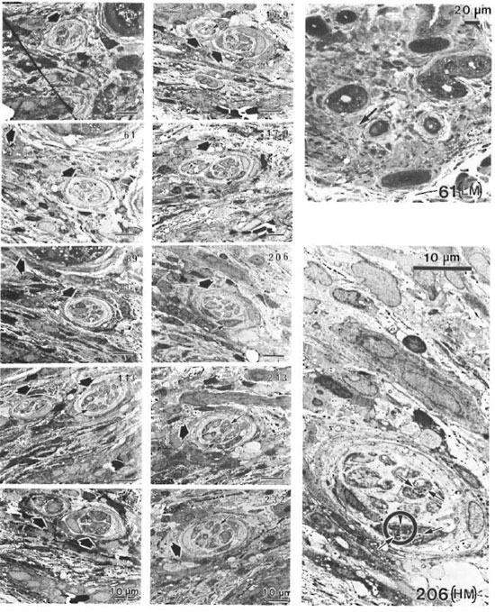
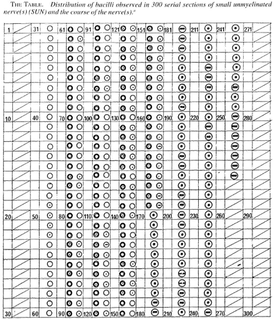
The course of this small nerve was traced through the neighboring serial sections, and the observations are summarized in The Table. Ten photomicrographs were selected and are presented in The Figure. A few bacilli were observed in vacuolar spaces located in the axoplasm (← in photos 113,169, 170, 206, 213, 232, and 206 [HM] in The Figure). They were diverse and some were seen in the act of dividing ( in photo 206 [HM]). We also saw two nerves joining to form one nerve, or vice versa, as seen in photo 170 in The Table and
  in photos 169 and 170 in The Figure.
In the literature it has been emphasized that intra-axonal leprosy bacilli were only to be found in myelinated axons and that none were to be seen ¡11 unmyelinated axons (1,3,4). The present observations show thai bacilli can he found in unmyelinated axons. Harlier studies, based on the usual electron microscopic examinations, would not have been likely to observe such bacilli since the usual studies do not employ serial sections.
The locations of Mycobacterium leprae in dermal peripheral nerves in leprosy patients should be reinvestigated.

The figure . Ten photomicrographs of small unmyelinated nerve (s) (SUN) selected from photographs of 300 serial semithin sections published earlier (2).  = Small unmyelinated nerve; ← = a few bacillary cells observed in vacuolar spaces located in axoplasm; 61 [LM] = low magnification photomicrograph of 61 with (←) a few bacillary cells observed in vacuolar spaces located in axoplasm; 206 [ HM ] = high magnification photomicrograph of 206 with (
 ) a bacillary cell caught in the act of division.
a In photos 1-30 and 257-300, no small unmyelinated nerves seen. In photos 31-60, one small unmyelinated nerve can he seen. In photos 61-169, two small unmyelinated nerves can he seen. In photo 170, one small unmyelinated nerve showing joining of two nerves into one nerve can be seen. In photos 171-260, one small un-myelinated nerve can he seen.
b  = Section did not show any small unmyelinated nerves;
  = small unmyelinated nerve (SUN);
  = SUN observed containing. cross sections of bacillary cells;
  = SUN observed containing longitudinaland cross sections of bacillary cells;
  = SUN observed containing bacillary cell in the act of division.
- Tsunchiko Hirata. Ph.D.
Chief, Research Training Section and Electron Microscopic Laboratory
National Institute for Leprosy Research
4-2-1 Aoba-cho
Higashimurayantashi
Tokyo 189, Japan
- Nobuo Harada, M.D., Ph.D.
Honorary Director
National Leprosarium Oku Khornyo-En
Okayama, Japan
REFERENCES
1. BODDINGIUS, J. The occurrence of Mycobacterium leprae within axons of peripheral nerves. Acta Neuropalhol. (Berl.) 27(1974)257-270.
2. HIRATA, T. and HARADA, N. Electron microscopic observations of the relationship between peripheral nerve tissue proper and an endoneurial capillary in a dermal lesion of a relapsed lepromalous patient. Int. J. Lepr. 62(1994)89-98.
3. JACOB, J. M.. SHETTY, V. P. and ANTIA, N. H. A morphological study of nerve biopsies from cases of multibacillary leprosy given multidrug therapy. Acta Neuropalhol. (Berl.) 85(t993)533-541.
4. SHETTY, V. P., ANTIA, N. H. and JACOB, J. M . The pathology of early leprous neuropathy. J. Neurol. Sci. 88(1988)115-131.
Reprint requests to Dr. Hirata at his present address: JRDC Senior Researcher (Leprology), Raj-Pracha-Samasai Institute. Phra-Pradaeng, Samutprakarn 10130,Thailand.
