- Volume 61 , Number 4
- Page: 634–5
An investigation of mast cells in two basic leprosy groups
To the Editor:
Leprosy is a spectral disease, the clinical and histopathological features of which depend upon the level of the cellular immune response to Mycobacterium leprae (7,8). The immunopathogenesis of leprosy skin lesions has not been clarified completely. It is, however, believed that several cell populations may participate. The aim of this study was to investigate the role of mast cells in the histopathogenesis of leprosy.
The mast cell population in skin specimens from 28 untreated Greek leprosy patients was investigated. The patients initially were classified according to the criteria of Ridley and Jopling (8). Two basic groups were studied, tuberculoid and lepromatous. The first group comprised 9 cases (2 tuberculoid, TT; 7 borderline tuberculoid, BT) and the second one, 19 cases (4 borderline lepromatous, BL; 15 lepromatous, LL). Four out of 15 LL cases had features of histoid leprosy (HL). The chloracetate esterase (Fast Blue RR) method in paraffin sections was used for the detection of mast cells (7).
The mast cell count was done by light microscopy using the following scale: + was given for 0-50 cells, ++ for 50-120 cells, + + + for 120-200 cells and + + + + for > 200 cells. Ten high-power fields were studied in each specimen.
The results are presented in The Table. The mast cell count in the first group was lower than that in the second (Figs. 1 and 2). This difference was found to be statistically significant (p < 0.001) according to the Mann-Whitney rank sum test. A lower number of mast cells also was observed in the borderline forms of leprosy. The distribution of mast cells in the tissues of the two groups was similar. Mast cells also were found under the epidermis in the Grenz zone in the majority of the lepromatous cases.
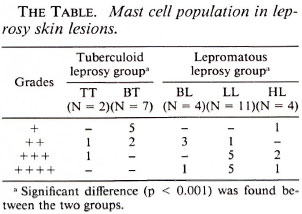
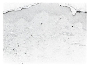
Fig. 1. Borderline tuberculoid leprosy, showing few scattered mast cells in the dermis (Fast Blue RR x250).
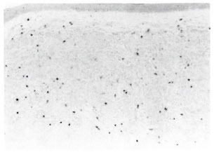
Fig. 2. Lepromatous leprosy, showing numerous mast cells (Fast Blue RR x250).
We did not note any important differences between the mast cell count in histoid leprosy specimens and those from other cases of the lepromatous group. We did, however, observe intensive degranulation in the former, a finding that reflects an increased functional activity and agrees with previous reports (3). Other researchers have noted the close adjacency of mast cells to lymphocytes and macrophages (9). Today it is believed that mast cells play an important role not only in immediate hypersensitivity reactions, but also in delayed hypersensitivity reactions (2), and that their functions are determined by factors deriving from both T lymphocytes and macrophages (10). Thus interleukin 3 (IL-3), IL-4, GM-CSF, and other mediators are involved in the growth, differentiation, and degranulation of mast cells, without being IgE-dependent, as probably occurs in histoid leprosy as well.
The predominance of mast cells in the lepromatous leprosy group as compared to the tuberculoid leprosy group also has been reported by other researchers (6,9). This increase in mast cells may be linked to the increased vascularity and changes observed in the endothelial cells, which are more obvious in lepromatous leprosy. Moreover, the intense presence and activity of mast cells in multibacillary leprosy could be due to the increased production of IL-3, IL-4, and GM-CSF by the CD8+ and Th2 population of CD4+ cells, which may be overactivated by M. leprae as has been proposed previously (5,11). The presence of mast cells in the Grenz zone, finally, might be related to various cytokines released from keratinocytes (4). Our findings reflect the variety of immunological reactions occurring in leprosy and imply a role for mast cells in these immunological phenomena.
- Kyriaki Aroni, M.D.
Associate Professor
Department of Pathology University of Athens
75 M. Asias str.
GR-115 27 Athens, Greece
- George Kontochristopoulos, M.D.
Hansen's Disease Center
General Hospital of Western Attica
Greece
- Anna Liossi, M.D.
Associate Professor
Department of Pathology
University of Athens
75 M. Asias str.
GR-115 27 Athens, Greece
- Dimitris Panteleos, M.D.
Hansen's Disease Center
General Hospital of Western Attica
Greece
REFERENCES
1. BANCROFT. .I. D. Enzyme histochemistry. In: Theory and Practice of Histological Techniques. 2nd ed. Bancroft, J. D. and Stevens, A., eds. Edinburgh: Churchill Livingstone. 1982, pp. 379-405.
2. GALLI, S. J. and DVORAK, A. M. What do mast cells have to do with delayed hypersensitivity? Lab. Invest. 50(1984)365-368.
3. KUMAR, R. Mast cells in histoid lepromatous lesions. Indian J. Lepr. 59(1987)390-392.
4. KUPPER, S. K. Mechanisms of cutaneous inflammation. Arch. Dermatol. 125(1989)1406-1412.
5. RAMOS, T., ZALCBERG-QUINTANA, I.. APPELBERG, R., SARNO, E. N. and SILVA. M . T . T-Helper subpopulations and the immune spectrum of leprosy. Int. J. Lepr. 57(1989)73-81.
6. RAV, S. D., PRATAP, V. K., SHARMA, N. K. and DAYAL, S. S. Mast cell in leprosy. Indian J. Lepr. 62(1990)467-472.
7. RIDLEY, D. S. Histological classification and the immunological spectrum of leprosy. Bull. WHO 51(1974)451-465.
8. RIDLEY, D. S. and JOPLING, W. H. Classification of leprosy according to immunity; a five-group system. Int. J. Lepr. 34(1966)255-273.
9. VAN HALL. H. M., TURKFL, S. B. and REA, T. H. Dermal ultrastructure in leprosy. Arch. Pathol. Lab. Med. 108(1984)383-386.
10. WASSERMAN. S. I. Mediators of inflammation. In: Basic and Clinical Immunology. 7th ed. Stites, D. P. and Terr, A. I., eds. Norwalk, Connecticut, U.S.A.: Appleton and Lange, 1991, pp. 154-160.
11. YAMAMURA, M.. UYEMURA, K., DEANS, R. J., WEINBERG, K., REA, T. H., BLOOM, B. R. and MODLIN. R. L. Defining protective responses to pathogens: cytokine profiles in leprosy lesions. Science 254(1991)277-279.