- Volume 68 , Number 1
- Page: 18–22
Comparative study of mitsuda reaction to nude mouse and armadillo lepromin preparations using nine-banded armadillos
ABSTRACT
In 14 nine-banded armadillos the Mitsuda response to nude mouse-derived lepromin (lepromin-nu/nu) was compared to that of armadillo-derived lepromin (lepromin-A) by injecting the reagents intradermally into either side of the abdomen of the animal and examining the biopsies f rom the sites after 12 days. The histopathologic responses to both antigens were found to be similar, whether the animal was Milsudanegative (lepromatous) or Mitsuda-positivc (tuberculoid). It is pointed out that armadillos arc good experimental models for leprosy, and their use can replace humans in experimental studies.RÉSUMÉ
La réponse de type Mitsuda, utilisant soit une lépromine dérivée de la souris nue (lépromine-nu/nu), soit une lépromine dérivée du tatou (lépromine A), fut comparée chez, le tatou à neuf bandes en injectant le produit dans le derme de chaque côté de l'abdomen de l'animal et en examinant les biopsies des sites d'injection après 12 jours. La réponse histopathologique contre chaque antigène fut jugée très semblable, que l'animal fût Mitsuda négatif (lépromateux) ou Mitsuda positif (tuberculoïde). Il est noté que les tatous sont des modèles expérimentaux très satisfaisants pour étudier la lèpre, et leur utilisation peut remplacer les humains dans les études expérimentales.RESUMEN
Se comparó la reacción de Mitsuda inducida con lepromina derivada de armadillo (lepromina A) con la inducida con lepromina derivada de ratón desnudo (lepromin-anu/nu) en 14 armadillos de nueve bandas. Las inyecciones se hicieron en los lados del abdomen y a los 12 días se tomaron biopsias de los sitios inyectados. El análisis histopatológico de las biopsias indicó que las respuestas hacia ambos antígenos fueron similares, independientemente de si los animales fueron Mitsuda-negativos (lepromatosos) o Mitsuda-positivos (tuberculoides). Se señala que los armadillos son un buen modelo experimental de la lepra y que su uso puede reemplazar a los humanos en ciertos estudios expérimentales.The Mitsuda reaction continues to be valuable for assessing the immune response to Mycobacterium leprae of leprosy patients and healthy individuals. Lepromatous nodules from patients with a high bacterial index (BI) used to be the source of M. leprae for preparing the lepromin reagent. Due to the reduction in the number of highly bacilliferous lepromatous patients worldwide, tissues harvested from lepromatous nine-banded armadillos (Dasypus novemcinctus) now have become the main source of M. leprae. Lepromin reagent prepared from infected armadillos (lepromin-A) has almost completely replaced lepromin of human origin (lcpromin-H). Now armadillos arc regularly used for the supply of bulk quantities of M. leprae.
Nude mice are the only other reliable source for M. leprae. The percentage of viable organisms in the nude mouse-derived samples of M. leprae is as high as or higher than that of specimens obtained from armadillos. Further, nude mouse-derived M. leprae can be readily mobilized in small quantities for daily routine use. Therefore, M. leprae derived from nude-mouse tissues are being used more and more in experimental studies. It was felt that a comparative study of the Mitsuda response of lepromin-A and lepromin of nude mouse origin (lepromin-nu/nu) in identical hosts would be in order.
MATERIALS AND METHODS
Preparation of lepromin reagents
Lepromin-A. All procedures were conducted under strict aseptic conditions. Lepromatous skin and subcutaneous nodules and lymph nodes from infected armadillos were collected and the fat was removed. The sterility of the tissues was tested using blood agar, thioglycollate and tripticase soy media. The tissues were autoclaved at 120ºC for 15 min, cut into small pieces, homogenized into a paste, suspended in saline (1 g of tissue for 10 ml of saline) and homogenized again. The suspension was then passed through a 70-µm nylon sterile/gamma-irradiated cell strainer. The strained suspension was tested for sterility as described above. Bacterial counts were made, and the suspension was diluted to contain 30 to 40 x 106 M. leprae per ml. Then 0.57 ml of 90% phenol per 100 ml was added. The suspension was then autoclaved at 120ºC for 15 min and cooled to room temperature. Before bottling, the final product was tested for safety according to the recommended guidelines of the World Health Organization (WHO) (11).
Lepromin-nu/nu. BALB nu/nu (Harlan, Sprague Dawley, Inc., Indianapolis, Indiana, U.S.A.) mice were injected in the hind foot pads with live M. leprae using the method described previously (1). Infected nude mice with enlarged foot pads were selected and sacrificed. The foot pads were exposed to ultraviolet irradiation for 3 min, washed with Betadine® for 20 min, and cleaned with 70% ethanol four times. The lepromatous nodules in the foot were dissected out by cutting out the epidermis and the underlying bones. The tissues thus obtained were processed exactly as described above for the armadillo tissues.
Lepromin testing
Fourteen armadillos caught from areas nonenzootic for leprosy and found negative for wild M. leprae infection were chosen for the study. One-tenth ml of lepromin-A was injected intradermally on one side of the abdomen and 0.1 ml of lepromin-nu/nu' on the opposite side of the abdomen. During injection, a small swelling was raised in the epidermis at the site of injection so as to be sure of the intradermal location of the injection. Lepromin bottles were shaken well before filling the syringes. The injection was made immediately so that the bacillary suspension was uniform. Separate syringes were used for each injection, and the site of injection was identified with tattoos.
At 21 days the test sites were biopsied with a 6-mm punch. The tissues were fixed in 10% neutral formalin, processed for paraffin sections, and 5-µm sections were cut. The sections were stained with hematoxylin and eosin (H&E) and with a modified Fite's stain for acid-fast bacilli (AFB). Since the armadillo skin is thick, gross readings of the reaction are unreliable and, therefore, were not taken.
Histopathology
The H&E reactions were studied and the granuloma fraction was assessed as a percentage of inflammatory cells replacing the dermis. The various types of cells, such as macrophages, epithelioid cells and lymphocytes, etc., were identified and described. The sections stained for AFB were assessed, and the bacterial load was graded from 1+ to 6+. The granuloma of the lepromin test was classified according to the criteria described earlier (6) into lepromatous (LL), borderline lepromatous (BL), borderline tuberculoid (BT) and tuberculoid (TT).
RESULTS
The summary of the results is given in The Table. The granuloma fraction occupying the dermis was about the same, except in 5 out of the 14 animals, and even so, the difference was only 5% to 10%. The cell types found in the granuloma showed no difference between these two groups (Figs. 1 a and b, 2 a and b). The bacterial load of the granuloma was the same in all except two animals. The biopsy was inadequate in one and, therefore, could not be properly assessed and in the other, the difference was 1 log. It should be noted here that in animals classified as BT, although the bacterial load was 0, in focal areas of necrosis small clumps of AFB were present. The classification of the Mitsuda response in both groups was the same. There were nine lcpromatous (LL) (Fig. 1 a and b) and five borderline tuberculoid (BT) (Fig. 2 a and b) animals. It was obvious that the Mitsuda responses in the armadillo to lepromin-A and lepromin-nu/nu were virtually identical.
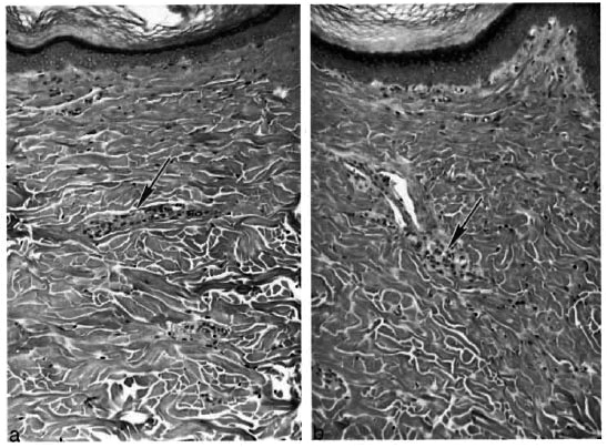
Fig. 1. Lepromin-negative armadillo (8-88) injected intradernially with 0.1 ml of lepromin-A. a In the dermis there were a few small collections of macrophages, one of which is shown in the photomicrograph (H&E X150). b Same animal (8-88) injected intradernially with 0.1 ml of lepromin-nu/nu. In the dermis there were a few granulomas almost identical in size and content, one of which is seen in the photomicrograph (H&E x150).
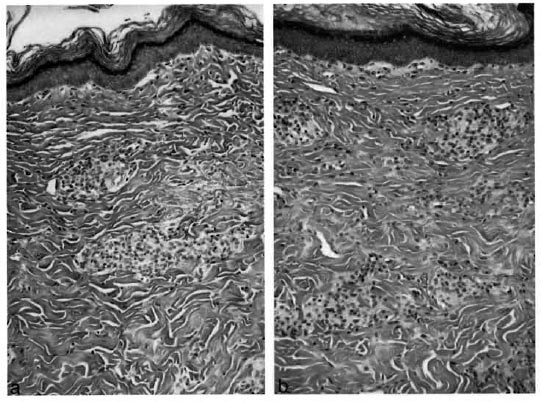
Fig. 2. Lepromin-positive armadillo (9-8) injected intradermally with 0.1 ml of lepromin-A. a Photomicrograph shows fairly large focal collections of epithelioid cells and lymphocytes in the dermis (H&E x 150).b Same animal (9-8) injected intradermally with 0.1 ml of lepromin-nu/nu. Photomicrograph shows granuloma slightly larger in size but the cellular contents were identical (H&Ex150).
DISCUSSION
Lepromin remains a useful index of the host's cell-mediated immune response to M. leprae, and is the most widely used skin-test preparation in leprosy. Comparative studies in human subjects of reactions to lepromin-H and lepromin-A have been reported (7- 9). The pattern of reactions evoked was the same with both lepromin reagents. In this study, lepromin-nu/nu and lepromin-A were found to produce identical Mitsuda responses in both Mitsuda-positive (Fig. 2 a and b) and Mitsuda-negative (Fig. 1 a and b) nine-banded armadillos. Instead of human subjects laboratory animals were used to demonstrate convincingly the comparability of lepromin-nu/nu and lepromin-A. This study again shows that the armadillo is a good and adequate animal for experimental studies and can, in many instances, take the place of expensive human experiments.
Integral lepromin is a twice-autoclaved extract of highly bacilliferous tissue. Although many proteins surely denature through such processing, antigens relevant to the host's granulomatous response obviously remain intact, and the 21-day Mitsuda reaction is considered to be specific to M. leprae. Draper, et al. showed that lepromins prepared with M. leprae derived from humans or mice gave equivalent reactions in man (3), and Meyers, et al. showed that even wild-type M. leprae obtained from naturally infected armadillos reacted identically in man as human-derived M. leprae propagated in armadillos (8). The host's granulomatous response to lepromin is influenced markedly by the concentration of M. leprae bacilli in the preparation (4- 5). However, no significant antigenic variation for M. leprae has been noted for lepromin, regardless of the geographic or host origin of the bacilli.
Since 1987 the GWL Hansen's Disease Center Laboratory has distributed 18 different batches of lepromin, each prepared with bacilli derived from different armadillos infected with different M. leprae isolates. No significant variation in reactivity has been noted between any of the batches prepared (unpublished observations). M. leprae appears to be highly conserved, and no significant genomic variation has been found between different isolates from different regions (2,10). The similarity seen in Mitsuda responses to different lepromins suggests that there is also relatively little variation in expression of antigens between different isolates or different propagative hosts, at least in regard to the major antigenic components influencing the host's granulomatous response to killed M. leprae.
Acknowledgment. We are grateful to Roena Domingue, Sumir Chehl, Erica Perrer and Joe Allen for technical help and to Penne Cason for secretarial assistance. The funds for this study were made available in part from NIAI1) Intra/agency Agreement #AI5015-01.
REFERENCES
1. CHEHL, S., Ruby, J., Job, C. K. and HASTINGS, R. C. The growth of M. leprae in nude mice. Lepr. Rev. 54(1983) 283-304.
2. DEWITT, M. Y. L. and KLATSER, P. R. M. leprae isolates from different sources have identical sequences of the spacer region between the 16S and 23S ribosomal RNA genes. Microbiology 140 (1994)1983-1987.
3. DRAPER, R., Rees. R. J. W. and WATERS, M. F. R. Comparison in man of lepromins prepared from leprosy infections in man and mice. Clin. Exp. Immunol. 3(1968) 809-816.
4. Hanks, J. H., Abe, M., Nakayma, T, Bechelu, L. M. and MARTINEZ, D. V. Studies toward the standardization of lepromin. Bull. WHO 42 (1970) 703-709.
5. Job , C. K., KlRCHHEIMER, W. F. and SANCHEZ, R. M. Variable lepromin response to Mycobacterium leprae in resistant armadillos. Int. J. Lepr. 51 (1983) 347-353.
6. Job, C. K., Sanchez, R. M., Hunt, R. and Hastings , R. C. Prevalence and significance of Mitsuda reaction in the nine-banded armadillo. Int. J. Lepr. 55(1987) 685-688.
7. Meyers, W. M., Kvermes, S. and Binford, C. II. Comparison of reactions to human and armadillo lepromin. Int. J. Lepr. 43 (1975) 218-225.
8. Meyers, w. M., Walsh, G. p., Binford, C. IL, STORRS, E. E. and Brown , H. L. Indigenous leprosy in nine-banded armadillos. In: The Armadillo as au Experimental Model in Biomedical Research. Vol. 366. Washington, DC: Pan American Health Organization, 1978, pp. 67-72.
9. Millar, J. W., Gannon, S. C. and Chan, C. S. P. Comparison in leprosy patients of Fernandez and Mitsuda reactions using human and armadillo antigens: a double-blind study. Int. J. Lepr. 43 (1975) 226-233.
10. Williams, D. L., Gillis, T. P. and Portaels, F. Geographically distinct isolates of M. leprae exhibit no genolypic diversity by restriction fragment polymorphism analysis. Mol. Microbiol. 4 (1990) 1653-1659.
11. World Health Organization. Recommended safety requirements for the preparation of lepromin: a WHO memorandum. Bull. WHO 57 (1979) 921-924.
1. M.D., Visiting Scientist; Microbiology Research Department, GWL Hansen's Disease Center at Louisiana State University, P.O. Box 25072, Baton Rouge, Louisiana 70894, U.S.A.
2. Visiting Scientist; Microbiology Research Department, GWL Hansen's Disease Center at Louisiana State University, P.O. Box 25072, Baton Rouge, Louisiana 70894, U.S.A.
3. Ph.D., Chief, Microbiology Research Department, GWL Hansen's Disease Center at Louisiana State University, P.O. Box 25072, Baton Rouge, Louisiana 70894, U.S.A.
Reprint requests to Dr. Truman at the above address or FAX: 1-225-346-5786.
Received for publication on 8 September 1999.
Accepted for publication on 13 October 1999.